5DO8

1.8 Angstrom crystal structure of Listeria monocytogenes Lmo0184 alpha-1,6-glucosidase


Experimental Data Snapshot

  • Method: X-RAY DIFFRACTION
  • Resolution: 1.80 Å
  • R-Value Free: 0.188 
  • R-Value Work: 0.154 
  • R-Value Observed: 0.155 

wwPDB Validation   3D Report Full Report


Ligand Structure Quality Assessment 


This is version 1.3 of the entry. See complete history


Literature

Structure to function of an alpha-glucan metabolic pathway that promotes Listeria monocytogenes pathogenesis.

Light, S.H.Cahoon, L.A.Halavaty, A.S.Freitag, N.E.Anderson, W.F.

(2016) Nat Microbiol 2: 16202-16202

  • DOI: https://doi.org/10.1038/nmicrobiol.2016.202
  • Primary Citation of Related Structures:  
    4KMQ, 5DO8, 5F7P, 5F7Q, 5F7R, 5F7S, 5F7U, 5F7V

  • PubMed Abstract: 

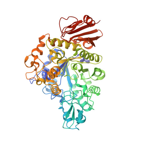
    Here we employ a 'systems structural biology' approach to functionally characterize an unconventional α-glucan metabolic pathway from the food-borne pathogen Listeria monocytogenes (Lm). Crystal structure determination coupled with basic biochemical and biophysical assays allowed for the identification of anabolic, transport, catabolic and regulatory portions of the cycloalternan pathway. These findings provide numerous insights into cycloalternan pathway function and reveal the mechanism of repressor, open reading frame, kinase (ROK) transcription regulators. Moreover, by developing a structural overview we were able to anticipate the cycloalternan pathway's role in the metabolism of partially hydrolysed starch derivatives and demonstrate its involvement in Lm pathogenesis. These findings suggest that the cycloalternan pathway plays a role in interspecies resource competition-potentially within the host gastrointestinal tract-and establish the methodological framework for characterizing bacterial systems of unknown function.


  • Organizational Affiliation

    Center for Structural Genomics of Infectious Diseases and Department of Biochemistry and Molecular Genetics, Feinberg School of Medicine, Northwestern University, Chicago, Illinois 60611, USA.


Macromolecules
Find similar proteins by:  (by identity cutoff)  |  3D Structure
Entity ID: 1
MoleculeChains Sequence LengthOrganismDetailsImage
Lmo0184 proteinA [auth B],
B [auth A],
C
555Listeria monocytogenes EGD-eMutation(s): 0 
Gene Names: lmo0184
UniProt
Find proteins for Q8YAE6 (Listeria monocytogenes serovar 1/2a (strain ATCC BAA-679 / EGD-e))
Explore Q8YAE6 
Go to UniProtKB:  Q8YAE6
Entity Groups  
Sequence Clusters30% Identity50% Identity70% Identity90% Identity95% Identity100% Identity
UniProt GroupQ8YAE6
Sequence Annotations
Expand
  • Reference Sequence
Small Molecules
Ligands 3 Unique
IDChains Name / Formula / InChI Key2D Diagram3D Interactions
BTB
Query on BTB

Download Ideal Coordinates CCD File 
F [auth B],
I [auth A],
M [auth C]
2-[BIS-(2-HYDROXY-ETHYL)-AMINO]-2-HYDROXYMETHYL-PROPANE-1,3-DIOL
C8 H19 N O5
OWMVSZAMULFTJU-UHFFFAOYSA-N
BGC
Query on BGC

Download Ideal Coordinates CCD File 
D [auth B],
G [auth A],
J [auth C]
beta-D-glucopyranose
C6 H12 O6
WQZGKKKJIJFFOK-VFUOTHLCSA-N
CL
Query on CL

Download Ideal Coordinates CCD File 
E [auth B],
H [auth A],
K [auth C],
L [auth C]
CHLORIDE ION
Cl
VEXZGXHMUGYJMC-UHFFFAOYSA-M
Experimental Data & Validation

Experimental Data

Unit Cell:
Length ( Å )Angle ( ˚ )
a = 88.123α = 90
b = 103.937β = 90
c = 193.79γ = 90
Software Package:
Software NamePurpose
REFMACrefinement
HKL-3000data reduction
HKL-3000data scaling
PHASERphasing

Structure Validation

View Full Validation Report


Ligand Structure Quality Assessment 


Entry History 

Deposition Data

Revision History  (Full details and data files)

  • Version 1.0: 2015-09-30
    Type: Initial release
  • Version 1.1: 2016-11-23
    Changes: Database references
  • Version 1.2: 2020-07-29
    Type: Remediation
    Reason: Carbohydrate remediation
    Changes: Data collection, Derived calculations, Structure summary
  • Version 1.3: 2023-09-27
    Changes: Data collection, Database references, Refinement description, Structure summary