5DNL

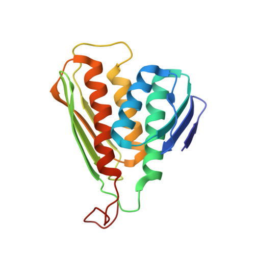
Crystal structure of IGPD from Pyrococcus furiosus in complex with (S)-C348


Experimental Data Snapshot

  • Method: X-RAY DIFFRACTION
  • Resolution: 1.53 Å
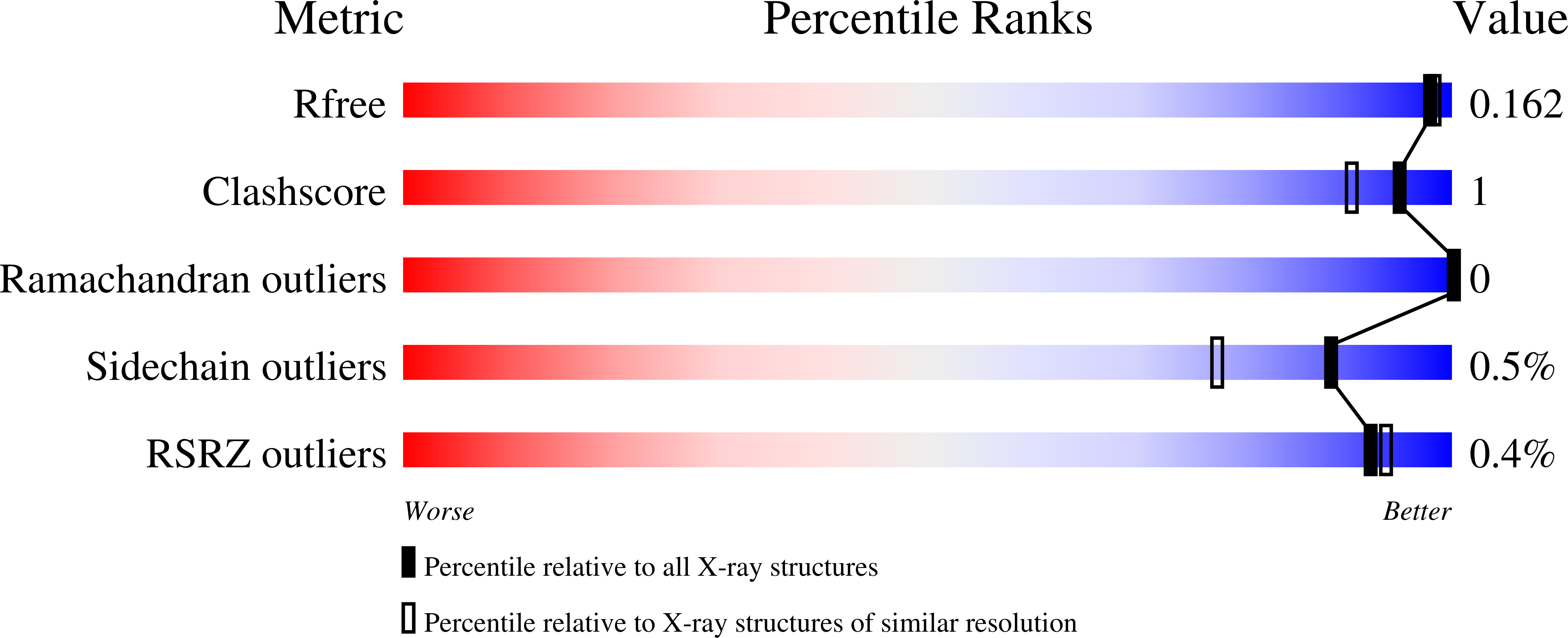
  • R-Value Free: 0.149 
  • R-Value Work: 0.135 
  • R-Value Observed: 0.136 

wwPDB Validation   3D Report Full Report


Ligand Structure Quality Assessment 


This is version 2.1 of the entry. See complete history


Literature

Mirror-Image Packing Provides a Molecular Basis for the Nanomolar Equipotency of Enantiomers of an Experimental Herbicide.

Bisson, C.Britton, K.L.Sedelnikova, S.E.Rodgers, H.F.Eadsforth, T.C.Viner, R.C.Hawkes, T.R.Baker, P.J.Rice, D.W.

(2016) Angew Chem Int Ed Engl 55: 13485-13489

  • DOI: https://doi.org/10.1002/anie.201607185
  • Primary Citation of Related Structures:  
    5DNL, 5DNX, 5EKW, 5EL9, 5ELW

  • PubMed Abstract: 

    Programs of drug discovery generally exploit one enantiomer of a chiral compound for lead development following the principle that enantiomer recognition is central to biological specificity. However, chiral promiscuity has been identified for a number of enzyme families, which have shown that mirror-image packing can enable opposite enantiomers to be accommodated in an enzyme's active site. Reported here is a series of crystallographic studies of complexes between an enzyme and a potent experimental herbicide whose chiral center forms an essential part of the inhibitor pharmacophore. Initial studies with a racemate at 1.85 Å resolution failed to identify the chirality of the bound inhibitor, however, by extending the resolution to 1.1 Å and by analyzing high-resolution complexes with the enantiopure compounds, we determined that both enantiomers make equivalent pseudosymmetric interactions in the active site, thus mimicking an achiral reaction intermediate.


  • Organizational Affiliation

    Krebs Institute for Biomolecular Research, Department of Molecular Biology and Biotechnology, University of Sheffield, Firth Court, Western Bank, Sheffield, S10 2TN, UK.


Macromolecules
Find similar proteins by:  (by identity cutoff)  |  3D Structure
Entity ID: 1
MoleculeChains Sequence LengthOrganismDetailsImage
Imidazoleglycerol-phosphate dehydrataseA,
B [auth C],
C [auth B]
176Pyrococcus furiosusMutation(s): 0 
Gene Names: hisBPF1660
EC: 4.2.1.19
UniProt
Find proteins for P58880 (Pyrococcus furiosus (strain ATCC 43587 / DSM 3638 / JCM 8422 / Vc1))
Explore P58880 
Go to UniProtKB:  P58880
Entity Groups  
Sequence Clusters30% Identity50% Identity70% Identity90% Identity95% Identity100% Identity
UniProt GroupP58880
Sequence Annotations
Expand
  • Reference Sequence
Small Molecules
Ligands 2 Unique
IDChains Name / Formula / InChI Key2D Diagram3D Interactions
5DL
Query on 5DL

Download Ideal Coordinates CCD File 
F [auth A],
I [auth C],
L [auth B]
[(2S)-2-hydroxy-3-(1H-1,2,4-triazol-1-yl)propyl]phosphonic acid
C5 H10 N3 O4 P
ZXKJPBBOMRHTCH-YFKPBYRVSA-N
MN
Query on MN

Download Ideal Coordinates CCD File 
D [auth A]
E [auth A]
G [auth C]
H [auth C]
J [auth B]
D [auth A],
E [auth A],
G [auth C],
H [auth C],
J [auth B],
K [auth B]
MANGANESE (II) ION
Mn
WAEMQWOKJMHJLA-UHFFFAOYSA-N
Experimental Data & Validation

Experimental Data

  • Method: X-RAY DIFFRACTION
  • Resolution: 1.53 Å
  • R-Value Free: 0.149 
  • R-Value Work: 0.135 
  • R-Value Observed: 0.136 
  • Space Group: I 4 2 2
Unit Cell:
Length ( Å )Angle ( ˚ )
a = 141.27α = 90
b = 141.27β = 90
c = 137.41γ = 90
Software Package:
Software NamePurpose
REFMACrefinement
XDSdata reduction
XSCALEdata scaling
PHASERphasing

Structure Validation

View Full Validation Report


Ligand Structure Quality Assessment 


Entry History & Funding Information

Deposition Data


Funding OrganizationLocationGrant Number
Biotechnology and Biological Sciences Research CouncilUnited Kingdom--

Revision History  (Full details and data files)

  • Version 1.0: 2016-09-28
    Type: Initial release
  • Version 1.1: 2016-10-05
    Changes: Database references
  • Version 1.2: 2016-10-19
    Changes: Database references
  • Version 1.3: 2016-10-26
    Changes: Database references
  • Version 2.0: 2017-08-30
    Changes: Advisory, Atomic model, Author supporting evidence, Derived calculations
  • Version 2.1: 2024-01-10
    Changes: Data collection, Database references, Derived calculations, Refinement description