5DNI

Crystal structure of Methanocaldococcus jannaschii Fumarate hydratase beta subunit


Experimental Data Snapshot

  • Method: X-RAY DIFFRACTION
  • Resolution: 2.30 Å
  • R-Value Free: 0.231 
  • R-Value Work: 0.181 
  • R-Value Observed: 0.183 

wwPDB Validation   3D Report Full Report


This is version 1.2 of the entry. See complete history


Literature

Revisiting the Burden Borne by Fumarase: Enzymatic Hydration of an Olefin.

Bellur, A.Das, S.Jayaraman, V.Behera, S.Suryavanshi, A.Balasubramanian, S.Balaram, P.Jindal, G.Balaram, H.

(2023) Biochemistry 62: 476-493

  • DOI: https://doi.org/10.1021/acs.biochem.2c00541
  • Primary Citation of Related Structures:  
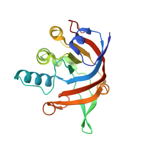
    5DNI, 7XKY

  • PubMed Abstract: 

    Fumarate hydratase (FH) is a remarkable catalyst that decreases the free energy of the catalyzed reaction by 30 kcal mol -1 , much larger than most exceptional enzymes with extraordinary catalytic rates. Two classes of FH are observed in nature: class-I and class-II, which have different folds, yet catalyze the same reversible hydration/dehydration reaction of the dicarboxylic acids fumarate/malate, with equal efficiencies. Using class-I FH from the hyperthermophilic archaeon Methanocaldococcus jannaschii (Mj) as a model along with comparative analysis with the only other available class-I FH structure from Leishmania major (Lm), we provide insights into the molecular mechanism of catalysis in this class of enzymes. The structure of MjFH apo-protein has been determined, revealing that large intersubunit rearrangements occur across apo- and holo-protein forms, with a largely preorganized active site for substrate binding. Site-directed mutagenesis of active site residues, kinetic analysis, and computational studies, including density functional theory (DFT) and natural population analysis, together show that residues interacting with the carboxylate group of the substrate play a pivotal role in catalysis. Our study establishes that an electrostatic network at the active site of class-I FH polarizes the substrate fumarate through interactions with its carboxylate groups, thereby permitting an easier addition of a water molecule across the olefinic bond. We propose a mechanism of catalysis in FH that occurs through transition-state stabilization involving the distortion of the electronic structure of the substrate olefinic bond mediated by the charge polarization of the bound substrate at the enzyme active site.


  • Organizational Affiliation

    Molecular Biology and Genetics Unit, Jawaharlal Nehru Centre for Advanced Scientific Research, Jakkur, Bengaluru 560064, India.


Macromolecules
Find similar proteins by:  (by identity cutoff)  |  3D Structure
Entity ID: 1
MoleculeChains Sequence LengthOrganismDetailsImage
Putative L(+)-tartrate dehydratase subunit beta
A, B
179Methanocaldococcus jannaschii DSM 2661Mutation(s): 0 
Gene Names: MjFHbetaMJ0617
EC: 4.2.1.32
UniProt
Find proteins for Q58034 (Methanocaldococcus jannaschii (strain ATCC 43067 / DSM 2661 / JAL-1 / JCM 10045 / NBRC 100440))
Explore Q58034 
Go to UniProtKB:  Q58034
Entity Groups  
Sequence Clusters30% Identity50% Identity70% Identity90% Identity95% Identity100% Identity
UniProt GroupQ58034
Sequence Annotations
Expand
  • Reference Sequence
Small Molecules
Ligands 4 Unique
IDChains Name / Formula / InChI Key2D Diagram3D Interactions
PEG
Query on PEG

Download Ideal Coordinates CCD File 
F [auth A]DI(HYDROXYETHYL)ETHER
C4 H10 O3
MTHSVFCYNBDYFN-UHFFFAOYSA-N
GOL
Query on GOL

Download Ideal Coordinates CCD File 
C [auth A],
D [auth A],
E [auth A],
H [auth B]
GLYCEROL
C3 H8 O3
PEDCQBHIVMGVHV-UHFFFAOYSA-N
EDO
Query on EDO

Download Ideal Coordinates CCD File 
I [auth B],
J [auth B],
K [auth B]
1,2-ETHANEDIOL
C2 H6 O2
LYCAIKOWRPUZTN-UHFFFAOYSA-N
CL
Query on CL

Download Ideal Coordinates CCD File 
G [auth A]CHLORIDE ION
Cl
VEXZGXHMUGYJMC-UHFFFAOYSA-M
Experimental Data & Validation

Experimental Data

  • Method: X-RAY DIFFRACTION
  • Resolution: 2.30 Å
  • R-Value Free: 0.231 
  • R-Value Work: 0.181 
  • R-Value Observed: 0.183 
  • Space Group: P 61
Unit Cell:
Length ( Å )Angle ( ˚ )
a = 85.61α = 90
b = 85.61β = 90
c = 121.83γ = 120
Software Package:
Software NamePurpose
PHENIXrefinement
MOSFLMdata collection
SCALAdata scaling
PDB_EXTRACTdata extraction
PHASERphasing
SCALAdata reduction

Structure Validation

View Full Validation Report


Entry History & Funding Information

Deposition Data


Funding OrganizationLocationGrant Number
Department of BiotechnologyIndia--

Revision History  (Full details and data files)

  • Version 1.0: 2016-09-14
    Type: Initial release
  • Version 1.1: 2023-02-08
    Changes: Database references, Derived calculations
  • Version 1.2: 2023-11-29
    Changes: Data collection, Refinement description