5DN4

Structure of the glycoside hydrolase domain from Salmonella typhimurium FlgJ


Experimental Data Snapshot

  • Method: X-RAY DIFFRACTION
  • Resolution: 1.80 Å
  • R-Value Free: 0.211 
  • R-Value Work: 0.182 
  • R-Value Observed: 0.183 

wwPDB Validation   3D Report Full Report


This is version 1.3 of the entry. See complete history


Literature

Structural and Biochemical Insights into the Peptidoglycan Hydrolase Domain of FlgJ from Salmonella typhimurium.

Zaloba, P.Bailey-Elkin, B.A.Derksen, M.Mark, B.L.

(2016) PLoS One 11: e0149204-e0149204

  • DOI: https://doi.org/10.1371/journal.pone.0149204
  • Primary Citation of Related Structures:  
    5DN4, 5DN5

  • PubMed Abstract: 

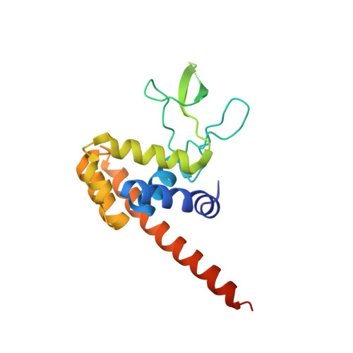
    FlgJ is a glycoside hydrolase (GH) enzyme belonging to the Carbohydrate Active enZyme (CAZy) family GH73. It facilitates passage of the bacterial flagellum through the peptidoglycan (PG) layer by cleaving the β-1,4 glycosidic bond between N-acetylglucosamine and N-acetylmuramic acid sugars that comprise the glycan strands of PG. Here we describe the crystal structure of the GH domain of FlgJ from bacterial pathogen Salmonella typhimurium (StFlgJ). Interestingly, the active site of StFlgJ was blocked by the C-terminal α-helix of a neighbouring symmetry mate and a β-hairpin containing the putative catalytic glutamic acid residue Glu223 was poorly resolved and could not be completely modeled into the electron density, suggesting it is flexible. Previous reports have shown that the GH73 enzyme Auto from Listeria monocytogenes is inhibited by an N-terminal α-helix that may occlude the active site in similar fashion. To investigate if the C-terminus of StFlgJ inhibits GH activity, the glycolytic activity of StFlgJ was assessed with and without the C-terminal α-helix. The GH activity of StFlgJ was unaffected by the presence or absence of the α-helix, suggesting it is not involved in regulating activity. Removal of the C-terminal α-helix did, however, allow a crystal structure of the domain to be obtained where the flexible β-hairpin containing residue Glu223 was entirely resolved. The β-hairpin was positioned such that the active site groove was fully solvent-exposed, placing Glu223 nearly 21.6 Å away from the putative general acid/base residue Glu184, which is too far apart for these two residues to coordinate glycosidic bond hydrolysis. The mobile nature of the StFlgJ β-hairpin is consistent with structural studies of related GH73 enzymes, suggesting that a dynamic active site may be common to many GH73 enzymes, in which the active site opens to capture substrate and then closes to correctly orient active site residues for catalysis.


  • Organizational Affiliation

    Department of Microbiology, University of Manitoba, Winnipeg, Canada.


Macromolecules
Find similar proteins by:  (by identity cutoff)  |  3D Structure
Entity ID: 1
MoleculeChains Sequence LengthOrganismDetailsImage
Peptidoglycan hydrolase FlgJ175Salmonella enterica subsp. enterica serovar Typhimurium str. LT2Mutation(s): 0 
Gene Names: flgJfla FXflaZSTM1182
EC: 3.2.1
UniProt
Find proteins for P15931 (Salmonella typhimurium (strain LT2 / SGSC1412 / ATCC 700720))
Explore P15931 
Go to UniProtKB:  P15931
Entity Groups  
Sequence Clusters30% Identity50% Identity70% Identity90% Identity95% Identity100% Identity
UniProt GroupP15931
Sequence Annotations
Expand
  • Reference Sequence
Small Molecules
Ligands 2 Unique
IDChains Name / Formula / InChI Key2D Diagram3D Interactions
IOD
Query on IOD

Download Ideal Coordinates CCD File 
B [auth A]
C [auth A]
D [auth A]
E [auth A]
F [auth A]
B [auth A],
C [auth A],
D [auth A],
E [auth A],
F [auth A],
G [auth A],
H [auth A],
I [auth A],
J [auth A],
K [auth A],
L [auth A]
IODIDE ION
I
XMBWDFGMSWQBCA-UHFFFAOYSA-M
CL
Query on CL

Download Ideal Coordinates CCD File 
M [auth A]CHLORIDE ION
Cl
VEXZGXHMUGYJMC-UHFFFAOYSA-M
Experimental Data & Validation

Experimental Data

  • Method: X-RAY DIFFRACTION
  • Resolution: 1.80 Å
  • R-Value Free: 0.211 
  • R-Value Work: 0.182 
  • R-Value Observed: 0.183 
  • Space Group: P 21 21 21
Unit Cell:
Length ( Å )Angle ( ˚ )
a = 38.79α = 90
b = 43.63β = 90
c = 107.93γ = 90
Software Package:
Software NamePurpose
PHENIXrefinement
MOSFLMdata reduction
Aimlessdata scaling
PHENIXphasing

Structure Validation

View Full Validation Report


Entry History & Funding Information

Deposition Data


Funding OrganizationLocationGrant Number
Natural Sciences and Engineering Research Council (NSERC, Canada)CanadaRGPIN-2015-05310

Revision History  (Full details and data files)

  • Version 1.0: 2016-02-24
    Type: Initial release
  • Version 1.1: 2017-09-20
    Changes: Author supporting evidence, Derived calculations
  • Version 1.2: 2020-01-08
    Changes: Author supporting evidence
  • Version 1.3: 2024-03-06
    Changes: Data collection, Database references, Derived calculations