5DMZ

Structure of human Bub1 kinase domain phosphorylated at Ser969


Experimental Data Snapshot

  • Method: X-RAY DIFFRACTION
  • Resolution: 2.40 Å
  • R-Value Free: 0.245 
  • R-Value Work: 0.210 
  • R-Value Observed: 0.212 

wwPDB Validation   3D Report Full Report


Ligand Structure Quality Assessment 


This is version 1.3 of the entry. See complete history


Literature

Role of Intrinsic and Extrinsic Factors in the Regulation of the Mitotic Checkpoint Kinase Bub1.

Breit, C.Bange, T.Petrovic, A.Weir, J.R.Muller, F.Vogt, D.Musacchio, A.

(2015) PLoS One 10: e0144673-e0144673

  • DOI: https://doi.org/10.1371/journal.pone.0144673
  • Primary Citation of Related Structures:  
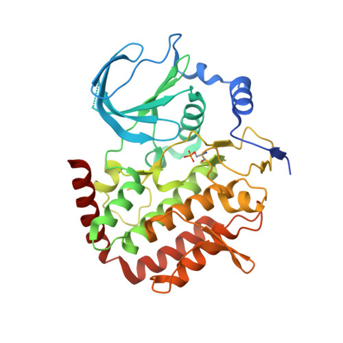
    5DMZ

  • PubMed Abstract: 

    The spindle assembly checkpoint (SAC) monitors microtubule attachment to kinetochores to ensure accurate sister chromatid segregation during mitosis. The SAC members Bub1 and BubR1 are paralogs that underwent significant functional specializations during evolution. We report an in-depth characterization of the kinase domains of Bub1 and BubR1. BubR1 kinase domain binds nucleotides but is unable to deliver catalytic activity in vitro. Conversely, Bub1 is an active kinase regulated by intra-molecular phosphorylation at the P+1 loop. The crystal structure of the phosphorylated Bub1 kinase domain illustrates a hitherto unknown conformation of the P+1 loop docked into the active site of the Bub1 kinase. Both Bub1 and BubR1 bind Bub3 constitutively. A hydrodynamic characterization of Bub1:Bub3 and BubR1:Bub3 demonstrates both complexes to have 1:1 stoichiometry, with no additional oligomerization. Conversely, Bub1:Bub3 and BubR1:Bub3 combine to form a heterotetramer. Neither BubR1:Bub3 nor Knl1, the kinetochore receptor of Bub1:Bub3, modulate the kinase activity of Bub1 in vitro, suggesting autonomous regulation of the Bub1 kinase domain. We complement our study with an analysis of the Bub1 substrates. Our results contribute to the mechanistic characterization of a crucial cell cycle checkpoint.


  • Organizational Affiliation

    Department of Mechanistic Cell Biology, Max Planck Institute of Molecular Physiology, Otto-Hahn-Straße 11, 44227, Dortmund, Germany.


Macromolecules
Find similar proteins by:  (by identity cutoff)  |  3D Structure
Entity ID: 1
MoleculeChains Sequence LengthOrganismDetailsImage
Mitotic checkpoint serine/threonine-protein kinase BUB1
A, B
365Homo sapiensMutation(s): 0 
Gene Names: BUB1BUB1L
EC: 2.7.11.1
UniProt & NIH Common Fund Data Resources
Find proteins for O43683 (Homo sapiens)
Explore O43683 
Go to UniProtKB:  O43683
PHAROS:  O43683
GTEx:  ENSG00000169679 
Entity Groups  
Sequence Clusters30% Identity50% Identity70% Identity90% Identity95% Identity100% Identity
UniProt GroupO43683
Sequence Annotations
Expand
  • Reference Sequence
Experimental Data & Validation

Experimental Data

  • Method: X-RAY DIFFRACTION
  • Resolution: 2.40 Å
  • R-Value Free: 0.245 
  • R-Value Work: 0.210 
  • R-Value Observed: 0.212 
  • Space Group: P 1 21 1
Unit Cell:
Length ( Å )Angle ( ˚ )
a = 62.38α = 90
b = 66.34β = 98.34
c = 106.54γ = 90
Software Package:
Software NamePurpose
PHENIXrefinement
XDSdata reduction
XSCALEdata scaling
PHASERphasing

Structure Validation

View Full Validation Report


Ligand Structure Quality Assessment 


Entry History & Funding Information

Deposition Data


Funding OrganizationLocationGrant Number
European Research Council (ERC) Advanced Investigator award RECEPIANCE--
German Research Foundation1093

Revision History  (Full details and data files)

  • Version 1.0: 2015-12-16
    Type: Initial release
  • Version 1.1: 2015-12-23
    Changes: Database references
  • Version 1.2: 2016-10-19
    Changes: Database references
  • Version 1.3: 2024-01-10
    Changes: Author supporting evidence, Data collection, Database references, Derived calculations, Refinement description