5DE2

Structural mechanism of Nek7 activation by Nek9-induced dimerisation


Experimental Data Snapshot

  • Method: X-RAY DIFFRACTION
  • Resolution: 2.78 Å
  • R-Value Free: 0.254 
  • R-Value Work: 0.206 
  • R-Value Observed: 0.208 

wwPDB Validation   3D Report Full Report


This is version 1.1 of the entry. See complete history


Literature

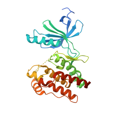
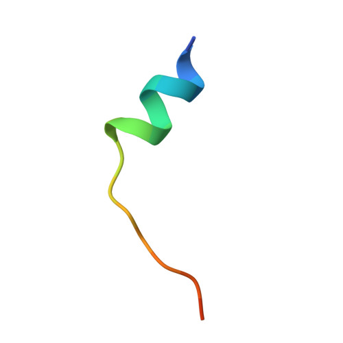
Mechanistic basis of Nek7 activation through Nek9 binding and induced dimerization.

Haq, T.Richards, M.W.Burgess, S.G.Gallego, P.Yeoh, S.O'Regan, L.Reverter, D.Roig, J.Fry, A.M.Bayliss, R.

(2015) Nat Commun 6: 8771-8771

  • DOI: https://doi.org/10.1038/ncomms9771
  • Primary Citation of Related Structures:  
    5DE2

  • PubMed Abstract: 

    Mitotic spindle assembly requires the regulated activities of protein kinases such as Nek7 and Nek9. Nek7 is autoinhibited by the protrusion of Tyr97 into the active site and activated by the Nek9 non-catalytic C-terminal domain (CTD). CTD binding apparently releases autoinhibition because mutation of Tyr97 to phenylalanine increases Nek7 activity independently of Nek9. Here we find that self-association of the Nek9-CTD is needed for Nek7 activation. We map the minimal Nek7 binding region of Nek9 to residues 810-828. A crystal structure of Nek7(Y97F) bound to Nek9(810-828) reveals a binding site on the C-lobe of the Nek7 kinase domain. Nek7(Y97F) crystallizes as a back-to-back dimer between kinase domain N-lobes, in which the specific contacts within the interface are coupled to the conformation of residue 97. Hence, we propose that the Nek9-CTD activates Nek7 through promoting back-to-back dimerization that releases the autoinhibitory tyrosine residue, a mechanism conserved in unrelated kinase families.


  • Organizational Affiliation

    Department of Biochemistry, University of Leicester, Lancaster Road, Leicester LE1 9HN, UK.


Macromolecules
Find similar proteins by:  (by identity cutoff)  |  3D Structure
Entity ID: 1
MoleculeChains Sequence LengthOrganismDetailsImage
Serine/threonine-protein kinase Nek7
A, B
310Homo sapiensMutation(s): 1 
Gene Names: NEK7
EC: 2.7.11.1
UniProt & NIH Common Fund Data Resources
Find proteins for Q8TDX7 (Homo sapiens)
Explore Q8TDX7 
Go to UniProtKB:  Q8TDX7
PHAROS:  Q8TDX7
GTEx:  ENSG00000151414 
Entity Groups  
Sequence Clusters30% Identity50% Identity70% Identity90% Identity95% Identity100% Identity
UniProt GroupQ8TDX7
Sequence Annotations
Expand
  • Reference Sequence

Find similar proteins by:  Sequence   |   3D Structure  

Entity ID: 2
MoleculeChains Sequence LengthOrganismDetailsImage
Serine/threonine-protein kinase Nek9
C, D
19Mus musculusMutation(s): 0 
EC: 2.7.11.1
UniProt & NIH Common Fund Data Resources
Find proteins for Q8K1R7 (Mus musculus)
Explore Q8K1R7 
Go to UniProtKB:  Q8K1R7
IMPC:  MGI:2387995
Entity Groups  
Sequence Clusters30% Identity50% Identity70% Identity90% Identity95% Identity100% Identity
UniProt GroupQ8K1R7
Sequence Annotations
Expand
  • Reference Sequence
Experimental Data & Validation

Experimental Data

  • Method: X-RAY DIFFRACTION
  • Resolution: 2.78 Å
  • R-Value Free: 0.254 
  • R-Value Work: 0.206 
  • R-Value Observed: 0.208 
  • Space Group: P 31 2 1
Unit Cell:
Length ( Å )Angle ( ˚ )
a = 88.12α = 90
b = 88.12β = 90
c = 155.61γ = 120
Software Package:
Software NamePurpose
BUSTERrefinement
XDSdata reduction
Aimlessdata scaling
PHASERphasing

Structure Validation

View Full Validation Report


Entry History & Funding Information

Deposition Data


Funding OrganizationLocationGrant Number
Cancer Research UKUnited KingdomC24461/ A12772

Revision History  (Full details and data files)

  • Version 1.0: 2015-11-11
    Type: Initial release
  • Version 1.1: 2024-01-10
    Changes: Data collection, Database references, Refinement description