5DCF

C-terminal domain of XerD recombinase in complex with gamma domain of FtsK


Experimental Data Snapshot

  • Method: X-RAY DIFFRACTION
  • Resolution: 2.30 Å
  • R-Value Free: 0.229 
  • R-Value Work: 0.191 
  • R-Value Observed: 0.193 

wwPDB Validation   3D Report Full Report


This is version 1.4 of the entry. See complete history


Literature

Activation of Xer-recombination at dif: structural basis of the FtsK gamma-XerD interaction.

Keller, A.N.Xin, Y.Boer, S.Reinhardt, J.Baker, R.Arciszewska, L.K.Lewis, P.J.Sherratt, D.J.Lowe, J.Grainge, I.

(2016) Sci Rep 6: 33357-33357

  • DOI: https://doi.org/10.1038/srep33357
  • Primary Citation of Related Structures:  
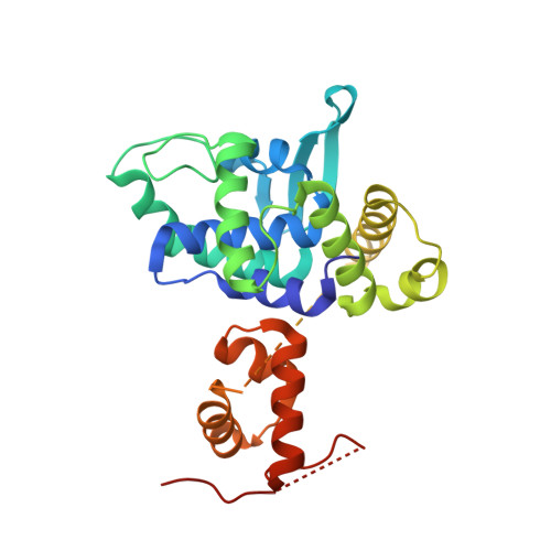
    5DCF

  • PubMed Abstract: 

    Bacterial chromosomes are most often circular DNA molecules. This can produce a topological problem; a genetic crossover from homologous recombination results in dimerization of the chromosome. A chromosome dimer is lethal unless resolved. A site-specific recombination system catalyses this dimer-resolution reaction at the chromosomal site dif. In Escherichia coli, two tyrosine-family recombinases, XerC and XerD, bind to dif and carry out two pairs of sequential strand exchange reactions. However, what makes the reaction unique among site-specific recombination reactions is that the first step, XerD-mediated strand exchange, relies on interaction with the very C-terminus of the FtsK DNA translocase. FtsK is a powerful molecular motor that functions in cell division, co-ordinating division with clearing chromosomal DNA from the site of septation and also acts to position the dif sites for recombination. This is a model system for unlinking, separating and segregating large DNA molecules. Here we describe the molecular detail of the interaction between XerD and FtsK that leads to activation of recombination as deduced from a co-crystal structure, biochemical and in vivo experiments. FtsKγ interacts with the C-terminal domain of XerD, above a cleft where XerC is thought to bind. We present a model for activation of recombination based on structural data.


  • Organizational Affiliation

    School of Environmental and Life Sciences, University of Newcastle, University Drive, Callaghan NSW 2308, Australia.


Macromolecules
Find similar proteins by:  (by identity cutoff)  |  3D Structure
Entity ID: 1
MoleculeChains Sequence LengthOrganismDetailsImage
Tyrosine recombinase XerD,DNA translocase FtsK275Escherichia coli CFT073Escherichia coli K-12
This entity is chimeric
Mutation(s): 0 
Gene Names: xerDc3474ftsKb0890JW0873
UniProt
Find proteins for P0A8P6 (Escherichia coli (strain K12))
Explore P0A8P6 
Go to UniProtKB:  P0A8P6
Entity Groups  
Sequence Clusters30% Identity50% Identity70% Identity90% Identity95% Identity100% Identity
UniProt GroupP0A8P6
Sequence Annotations
Expand
  • Reference Sequence
Experimental Data & Validation

Experimental Data

  • Method: X-RAY DIFFRACTION
  • Resolution: 2.30 Å
  • R-Value Free: 0.229 
  • R-Value Work: 0.191 
  • R-Value Observed: 0.193 
  • Space Group: P 65
Unit Cell:
Length ( Å )Angle ( ˚ )
a = 83.451α = 90
b = 83.451β = 90
c = 88.672γ = 120
Software Package:
Software NamePurpose
Aimlessdata scaling
PHASERphasing
ARPmodel building
PHENIXrefinement
PDB_EXTRACTdata extraction

Structure Validation

View Full Validation Report


Entry History & Funding Information

Deposition Data


Funding OrganizationLocationGrant Number
National Health and Medical Research Council (NHMRC, Australia)AustraliaAPP1005697 to I. Grainge

Revision History  (Full details and data files)

  • Version 1.0: 2016-09-07
    Type: Initial release
  • Version 1.1: 2016-11-16
    Changes: Database references
  • Version 1.2: 2017-09-20
    Changes: Author supporting evidence, Data collection, Derived calculations, Structure summary
  • Version 1.3: 2020-01-08
    Changes: Author supporting evidence
  • Version 1.4: 2023-09-27
    Changes: Data collection, Database references, Refinement description