5DBY

Crystal Structure of Equine Serum Albumin in Complex with Diclofenac and Naproxen Obtained in Displacement Experiment


Experimental Data Snapshot

  • Method: X-RAY DIFFRACTION
  • Resolution: 2.35 Å
  • R-Value Free: 0.234 
  • R-Value Work: 0.193 
  • R-Value Observed: 0.195 

wwPDB Validation   3D Report Full Report


Ligand Structure Quality Assessment 


This is version 1.2 of the entry. See complete history


Literature

Structural Insights into the Competitive Binding of Diclofenac and Naproxen by Equine Serum Albumin.

Sekula, B.Bujacz, A.

(2016) J Med Chem 59: 82-89

  • DOI: https://doi.org/10.1021/acs.jmedchem.5b00909
  • Primary Citation of Related Structures:  
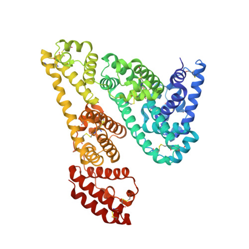
    4ZBQ, 4ZBR, 5DBY

  • PubMed Abstract: 

    The binding modes to equine serum albumin (ESA) of two nonsteroidal anti-inflammatory drugs (NSAIDs), diclofenac (Dic) and naproxen (Nps), were studied by X-ray crystallography and isothermal titration calorimetry. On the basis of the crystal structure of ESA/Dic determined to a resolution of 1.92 Å and the structure of the previously described ESA/Nps complex (2.42 Å), it was found that both NSAIDs bind within drug site 2 (DS2) of ESA and both occupy secondary binding sites in separate cavities of domain II (Nps) and domain III (Dic). The two structures of the ternary complex ESA/Dic/Nps, obtained by competitive cocrystallization (2.19 Å) and through a displacement experiment (2.35 Å), were determined to investigate possible competition of these widely used pharmaceutical drugs in binding to ESA. In these complexes Nps occupies the DS2 pocket common for both drugs, whereas the other distinct binding sites of Dic and Nps remain unaffected. These results suggest that combined application of both drugs may result in increased concentration of free diclofenac in plasma.


  • Organizational Affiliation

    Institute of Technical Biochemistry, Faculty of Biotechnology and Food Sciences, Lodz University of Technology , Stefanowskiego 4/10, 90-924 Lodz, Poland.


Macromolecules
Find similar proteins by:  (by identity cutoff)  |  3D Structure
Entity ID: 1
MoleculeChains Sequence LengthOrganismDetailsImage
Serum albumin583Equus caballusMutation(s): 0 
UniProt
Find proteins for P35747 (Equus caballus)
Explore P35747 
Go to UniProtKB:  P35747
Entity Groups  
Sequence Clusters30% Identity50% Identity70% Identity90% Identity95% Identity100% Identity
UniProt GroupP35747
Sequence Annotations
Expand
  • Reference Sequence
Small Molecules
Ligands 7 Unique
IDChains Name / Formula / InChI Key2D Diagram3D Interactions
DIF
Query on DIF

Download Ideal Coordinates CCD File 
B [auth A]2-[2,6-DICHLOROPHENYL)AMINO]BENZENEACETIC ACID
C14 H11 Cl2 N O2
DCOPUUMXTXDBNB-UHFFFAOYSA-N
NPS
Query on NPS

Download Ideal Coordinates CCD File 
C [auth A](2S)-2-(6-methoxynaphthalen-2-yl)propanoic acid
C14 H14 O3
CMWTZPSULFXXJA-VIFPVBQESA-N
LMR
Query on LMR

Download Ideal Coordinates CCD File 
Q [auth A](2S)-2-hydroxybutanedioic acid
C4 H6 O5
BJEPYKJPYRNKOW-REOHCLBHSA-N
SIN
Query on SIN

Download Ideal Coordinates CCD File 
I [auth A]SUCCINIC ACID
C4 H6 O4
KDYFGRWQOYBRFD-UHFFFAOYSA-N
MLI
Query on MLI

Download Ideal Coordinates CCD File 
D [auth A],
E [auth A],
F [auth A],
G [auth A],
H [auth A]
MALONATE ION
C3 H2 O4
OFOBLEOULBTSOW-UHFFFAOYSA-L
ACT
Query on ACT

Download Ideal Coordinates CCD File 
J [auth A]
K [auth A]
L [auth A]
M [auth A]
N [auth A]
J [auth A],
K [auth A],
L [auth A],
M [auth A],
N [auth A],
R [auth A]
ACETATE ION
C2 H3 O2
QTBSBXVTEAMEQO-UHFFFAOYSA-M
FMT
Query on FMT

Download Ideal Coordinates CCD File 
O [auth A],
P [auth A]
FORMIC ACID
C H2 O2
BDAGIHXWWSANSR-UHFFFAOYSA-N
Binding Affinity Annotations 
IDSourceBinding Affinity
DIF Binding MOAD:  5DBY Ka: 3.90e+4 (M^-1) from 1 assay(s)
NPS Binding MOAD:  5DBY Ka: 7.73e+6 (M^-1) from 1 assay(s)
Experimental Data & Validation

Experimental Data

  • Method: X-RAY DIFFRACTION
  • Resolution: 2.35 Å
  • R-Value Free: 0.234 
  • R-Value Work: 0.193 
  • R-Value Observed: 0.195 
  • Space Group: P 61
Unit Cell:
Length ( Å )Angle ( ˚ )
a = 94.32α = 90
b = 94.32β = 90
c = 141.69γ = 120
Software Package:
Software NamePurpose
REFMACrefinement
REFMACphasing
XSCALEdata scaling
XDSdata reduction

Structure Validation

View Full Validation Report


Ligand Structure Quality Assessment 


Entry History & Funding Information

Deposition Data


Funding OrganizationLocationGrant Number
Polsih Ministry of Science and Higher EducationPolandN N405 3636 39
National Science Centre PolandPoland2013/11/B/ST5/02271
National Science Centre PolandPoland2014/12/T/ST5/00136

Revision History  (Full details and data files)

  • Version 1.0: 2015-12-23
    Type: Initial release
  • Version 1.1: 2016-01-27
    Changes: Database references
  • Version 1.2: 2024-01-10
    Changes: Data collection, Database references, Refinement description