5DBJ

Crystal structure of halogenase PltA


Experimental Data Snapshot

  • Method: X-RAY DIFFRACTION
  • Resolution: 2.75 Å
  • R-Value Free: 0.292 
  • R-Value Work: 0.239 
  • R-Value Observed: 0.241 

wwPDB Validation   3D Report Full Report


Ligand Structure Quality Assessment 


This is version 1.3 of the entry. See complete history


Literature

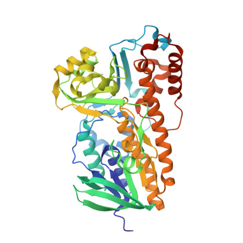
Crystal structure of halogenase PltA from the pyoluteorin biosynthetic pathway.

Pang, A.H.Garneau-Tsodikova, S.Tsodikov, O.V.

(2015) J Struct Biol 192: 349-357

  • DOI: https://doi.org/10.1016/j.jsb.2015.09.013
  • Primary Citation of Related Structures:  
    5DBJ

  • PubMed Abstract: 

    Pyoluteorin is an antifungal agent composed of a 4,5-dichlorinated pyrrole group linked to a resorcinol moiety. The pyoluteorin biosynthetic gene cluster in Pseudomonas fluorescens Pf-5 encodes the halogenase PltA, which has been previously demonstrated to perform both chlorinations in vitro. PltA selectively accepts as a substrate a pyrrole moiety covalently tethered to a nonribosomal peptide thiolation domain PltL (pyrrolyl-S-PltL) for FAD-dependent di-chlorination, yielding 4,5-dichloropyrrolyl-S-PltL. We report a 2.75 Å-resolution crystal structure of PltA in complex with FAD and chloride. PltA is a dimeric enzyme, containing a flavin-binding fold conserved in flavin-dependent halogenases and monooxygenases, and an additional unique helical region at the C-terminus. This C-terminal region blocks a putative substrate-binding cleft, suggesting that a conformational change involving repositioning of this region is necessary to allow binding of the pyrrolyl-S-PltL substrate for its dichlorination by PltA.


  • Organizational Affiliation

    Department of Pharmaceutical Sciences, College of Pharmacy, University of Kentucky, 789 South Limestone Street, Lexington, KY 40536-0596, USA.


Macromolecules
Find similar proteins by:  (by identity cutoff)  |  3D Structure
Entity ID: 1
MoleculeChains Sequence LengthOrganismDetailsImage
FADH2-dependent halogenase PltAA [auth E],
B [auth A],
C [auth B],
D [auth C],
E [auth D]
455Pseudomonas protegens Pf-5Mutation(s): 0 
Gene Names: pltAPFL_2787
EC: 3.8.1.1
UniProt
Find proteins for Q4KCZ0 (Pseudomonas fluorescens (strain ATCC BAA-477 / NRRL B-23932 / Pf-5))
Explore Q4KCZ0 
Go to UniProtKB:  Q4KCZ0
Entity Groups  
Sequence Clusters30% Identity50% Identity70% Identity90% Identity95% Identity100% Identity
UniProt GroupQ4KCZ0
Sequence Annotations
Expand
  • Reference Sequence
Small Molecules
Ligands 2 Unique
IDChains Name / Formula / InChI Key2D Diagram3D Interactions
FAD
Query on FAD

Download Ideal Coordinates CCD File 
F [auth E],
I [auth A],
L [auth B],
O [auth C],
R [auth D]
FLAVIN-ADENINE DINUCLEOTIDE
C27 H33 N9 O15 P2
VWWQXMAJTJZDQX-UYBVJOGSSA-N
CL
Query on CL

Download Ideal Coordinates CCD File 
G [auth E]
H [auth E]
J [auth A]
K [auth A]
M [auth B]
G [auth E],
H [auth E],
J [auth A],
K [auth A],
M [auth B],
N [auth B],
P [auth C],
Q [auth C],
S [auth D]
CHLORIDE ION
Cl
VEXZGXHMUGYJMC-UHFFFAOYSA-M
Experimental Data & Validation

Experimental Data

  • Method: X-RAY DIFFRACTION
  • Resolution: 2.75 Å
  • R-Value Free: 0.292 
  • R-Value Work: 0.239 
  • R-Value Observed: 0.241 
  • Space Group: C 1 2 1
Unit Cell:
Length ( Å )Angle ( ˚ )
a = 242.934α = 90
b = 94.981β = 91.21
c = 102.145γ = 90
Software Package:
Software NamePurpose
REFMACrefinement
HKL-2000data reduction
HKL-2000data scaling
PHASERphasing

Structure Validation

View Full Validation Report


Ligand Structure Quality Assessment 


Entry History 

Deposition Data

Revision History  (Full details and data files)

  • Version 1.0: 2015-10-07
    Type: Initial release
  • Version 1.1: 2015-10-14
    Changes: Database references
  • Version 1.2: 2015-11-25
    Changes: Database references
  • Version 1.3: 2023-09-27
    Changes: Data collection, Database references, Derived calculations, Refinement description