5D8L

Human HSF2 DNA Binding Domain in complex with 3-site HSE DNA at 2.1 Angstroms Resolution


Experimental Data Snapshot

  • Method: X-RAY DIFFRACTION
  • Resolution: 2.07 Å
  • R-Value Free: 0.273 
  • R-Value Work: 0.217 
  • R-Value Observed: 0.220 

wwPDB Validation   3D Report Full Report


This is version 1.4 of the entry. See complete history


Literature

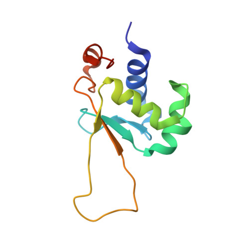
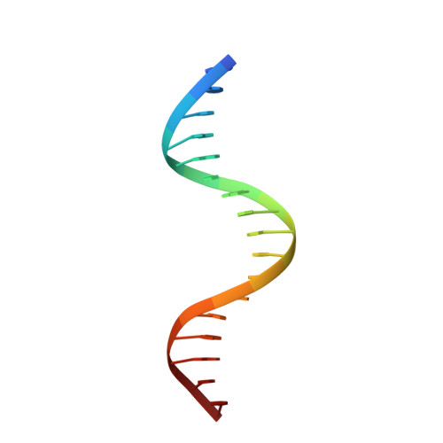
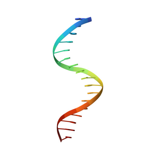
Structures of HSF2 reveal mechanisms for differential regulation of human heat-shock factors.

Jaeger, A.M.Pemble, C.W.Sistonen, L.Thiele, D.J.

(2016) Nat Struct Mol Biol 23: 147-154

  • DOI: https://doi.org/10.1038/nsmb.3150
  • Primary Citation of Related Structures:  
    5D8K, 5D8L

  • PubMed Abstract: 

    Heat-shock transcription factor (HSF) family members function in stress protection and in human diseases including proteopathies, neurodegeneration and cancer. The mechanisms that drive distinct post-translational modifications, cofactor recruitment and target-gene activation for specific HSF paralogs are unknown. We present crystal structures of the human HSF2 DNA-binding domain (DBD) bound to DNA, revealing an unprecedented view of HSFs that provides insights into their unique biology. The HSF2 DBD structures resolve a new C-terminal helix that directs wrapping of the coiled-coil domain around DNA, thereby exposing paralog-specific sequences of the DBD surface for differential post-translational modifications and cofactor interactions. We further demonstrate a direct interaction between HSF1 and HSF2 through their coiled-coil domains. Together, these features provide a new model for HSF structure as the basis for differential and combinatorial regulation, which influences the transcriptional response to cellular stress.


  • Organizational Affiliation

    Department of Pharmacology and Cancer Biology, Duke University School of Medicine, Durham, North Carolina, USA.


Macromolecules

Find similar proteins by:  (by identity cutoff)  |  3D Structure
Entity ID: 2
MoleculeChains Sequence LengthOrganismDetailsImage
Heat shock factor protein 2
B, D, F, H
110Homo sapiensMutation(s): 0 
Gene Names: HSF2HSTF2
UniProt & NIH Common Fund Data Resources
Find proteins for Q03933 (Homo sapiens)
Explore Q03933 
Go to UniProtKB:  Q03933
PHAROS:  Q03933
GTEx:  ENSG00000025156 
Entity Groups  
Sequence Clusters30% Identity50% Identity70% Identity90% Identity95% Identity100% Identity
UniProt GroupQ03933
Sequence Annotations
Expand
  • Reference Sequence

Find similar nucleic acids by:  Sequence   |   3D Structure  

Entity ID: 1
MoleculeChains LengthOrganismImage
DNA (5'-D(P*GP*TP*GP*AP*AP*TP*AP*TP*TP*CP*TP*AP*GP*AP*AP*CP*C)-3')
A, G
17synthetic construct
Sequence Annotations
Expand
  • Reference Sequence

Find similar nucleic acids by:  Sequence   |   3D Structure  

Entity ID: 3
MoleculeChains LengthOrganismImage
DNA (5'-D(*GP*GP*TP*TP*CP*TP*AP*GP*AP*AP*TP*AP*TP*TP*CP*AP*C)-3')
C, E
17synthetic construct
Sequence Annotations
Expand
  • Reference Sequence
Experimental Data & Validation

Experimental Data

  • Method: X-RAY DIFFRACTION
  • Resolution: 2.07 Å
  • R-Value Free: 0.273 
  • R-Value Work: 0.217 
  • R-Value Observed: 0.220 
  • Space Group: P 1
Unit Cell:
Length ( Å )Angle ( ˚ )
a = 40.174α = 82.82
b = 47.14β = 90.03
c = 109.587γ = 64.75
Software Package:
Software NamePurpose
PHENIXrefinement
HKL-2000data reduction
HKL-2000data scaling
PHASERphasing

Structure Validation

View Full Validation Report


Entry History 

Deposition Data

Revision History  (Full details and data files)

  • Version 1.0: 2016-01-06
    Type: Initial release
  • Version 1.1: 2016-02-03
    Changes: Database references
  • Version 1.2: 2016-02-10
    Changes: Database references
  • Version 1.3: 2017-09-27
    Changes: Author supporting evidence, Derived calculations
  • Version 1.4: 2024-03-06
    Changes: Data collection, Database references