5D7W

Crystal structure of serralysin


Experimental Data Snapshot

  • Method: X-RAY DIFFRACTION
  • Resolution: 1.10 Å
  • R-Value Free: 0.186 
  • R-Value Work: 0.175 
  • R-Value Observed: 0.176 

wwPDB Validation   3D Report Full Report


This is version 1.2 of the entry. See complete history


Literature

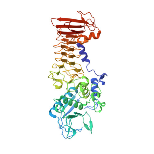
Structure of a thermostable serralysin from Serratia sp. FS14 at 1.1 angstrom resolution.

Wu, D.Ran, T.Wang, W.Xu, D.

(2016) Acta Crystallogr F Struct Biol Commun 72: 10-15

  • DOI: https://doi.org/10.1107/S2053230X15023092
  • Primary Citation of Related Structures:  
    5D7W

  • PubMed Abstract: 

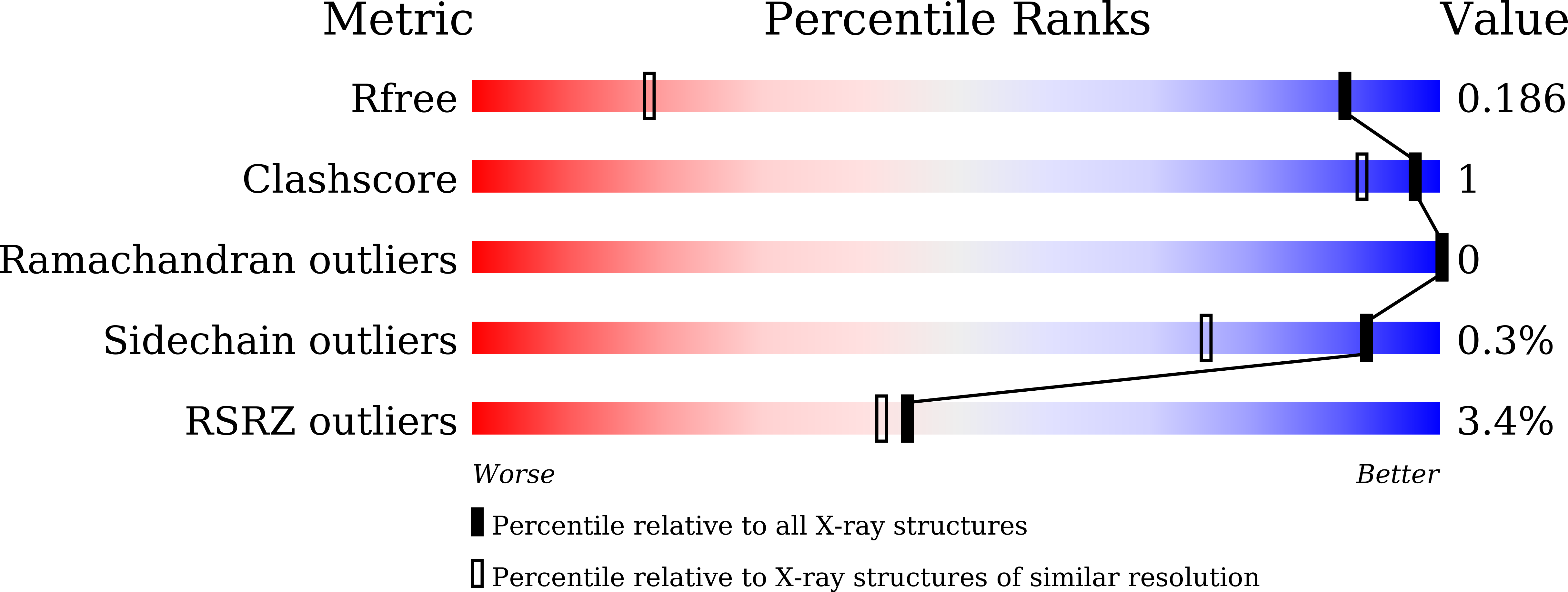
    Serralysin is a well studied metalloprotease, and typical serralysins are not thermostable. The serralysin isolated from Serratia sp. FS14 was found to be thermostable, and in order to reveal the mechanism responsible for its thermostability, the crystal structure of serralysin from Serratia sp. FS14 was solved to a crystallographic R factor of 0.1619 at 1.10 Å resolution. Similar to its homologues, it mainly consists of two domains: an N-terminal catalytic domain and a `parallel β-roll' C-terminal domain. Comparative studies show that the shape of the catalytic active-site cavity is more open owing to the 189-198 loop, with a short 310-helix protruding further from the molecular surface, and that the β-sheets comprising the `parallel β-roll' are longer than those in its homologues. The formation of hydrogen bonds from one of the nonconserved residues (Asn200) to Lys27 may contribute to the thermostability.


  • Organizational Affiliation

    Department of Microbiology, Nanjing Agricultural University, No. 1 Weigang, Nanjing, Jiangsu 210095, People's Republic of China.


Macromolecules
Find similar proteins by:  (by identity cutoff)  |  3D Structure
Entity ID: 1
MoleculeChains Sequence LengthOrganismDetailsImage
Serralysin469Serratia marcescensMutation(s): 1 
EC: 3.4.24.40
UniProt
Find proteins for P23694 (Serratia marcescens)
Explore P23694 
Go to UniProtKB:  P23694
Entity Groups  
Sequence Clusters30% Identity50% Identity70% Identity90% Identity95% Identity100% Identity
UniProt GroupP23694
Sequence Annotations
Expand
  • Reference Sequence
Small Molecules
Ligands 3 Unique
IDChains Name / Formula / InChI Key2D Diagram3D Interactions
GOL
Query on GOL

Download Ideal Coordinates CCD File 
J [auth A],
K [auth A]
GLYCEROL
C3 H8 O3
PEDCQBHIVMGVHV-UHFFFAOYSA-N
ZN
Query on ZN

Download Ideal Coordinates CCD File 
B [auth A]ZINC ION
Zn
PTFCDOFLOPIGGS-UHFFFAOYSA-N
CA
Query on CA

Download Ideal Coordinates CCD File 
C [auth A]
D [auth A]
E [auth A]
F [auth A]
G [auth A]
C [auth A],
D [auth A],
E [auth A],
F [auth A],
G [auth A],
H [auth A],
I [auth A]
CALCIUM ION
Ca
BHPQYMZQTOCNFJ-UHFFFAOYSA-N
Experimental Data & Validation

Experimental Data

  • Method: X-RAY DIFFRACTION
  • Resolution: 1.10 Å
  • R-Value Free: 0.186 
  • R-Value Work: 0.175 
  • R-Value Observed: 0.176 
  • Space Group: P 21 21 21
Unit Cell:
Length ( Å )Angle ( ˚ )
a = 42.3α = 90
b = 105.4β = 90
c = 151.1γ = 90
Software Package:
Software NamePurpose
PHENIXrefinement
XDSdata reduction
XSCALEdata scaling
PHASERphasing

Structure Validation

View Full Validation Report


Entry History & Funding Information

Deposition Data


Funding OrganizationLocationGrant Number
National Natural Science Foundation of ChinaChina31100028
National Natural Science Foundation of ChinaChina31400055
National Natural Science Foundation of ChinaChina31170686

Revision History  (Full details and data files)

  • Version 1.0: 2016-01-13
    Type: Initial release
  • Version 1.1: 2019-12-18
    Changes: Database references, Derived calculations, Structure summary
  • Version 1.2: 2023-11-08
    Changes: Data collection, Database references, Derived calculations, Refinement description