Enhancing Mn(II)-Binding and Manganese Peroxidase Activity in a Designed Cytochrome c Peroxidase through Fine-Tuning Secondary-Sphere Interactions.
Hosseinzadeh, P., Mirts, E.N., Pfister, T.D., Gao, Y.G., Mayne, C., Robinson, H., Tajkhorshid, E., Lu, Y.(2016) Biochemistry 55: 1494-1502
- PubMed: 26885726
- DOI: https://doi.org/10.1021/acs.biochem.5b01299
- Primary Citation of Related Structures:
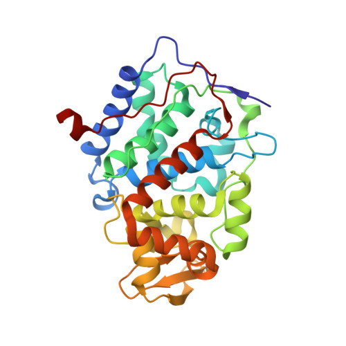
5D6M - PubMed Abstract:
Noncovalent second-shell interactions are important in controlling metal-binding affinity and activity in metalloenzymes, but fine-tuning these interactions in designed metalloenzymes has not been fully explored. As a result, most designed metalloenzymes have low metal-binding affinity and activity. Here we identified three mutations in the second coordination shell of an engineered Mn(II)-binding site in cytochrome c peroxidase (called MnCcP.1, containing Glu45, Glu37, and Glu181 ligands) that mimics the native manganese peroxidase (MnP), and explored their effects on both Mn(II)-binding affinity and MnP activity. First, removing a hydrogen bond to Glu45 through Tyr36Phe mutation enhanced Mn(II)-binding affinity, as evidenced by a 2.8-fold decrease in the KM of Mn(II) oxidation. Second, introducing a salt bridge through Lys179Arg mutation improved Glu35 and Glu181 coordination to Mn(II), decreasing KM 2.6-fold. Third, eliminating a steric clash that prevented Glu37 from orienting toward Mn(II) resulted in an 8.6-fold increase in kcat/KM, arising primarily from a 3.6-fold decrease in KM, with a KM value comparable to that of the native enzyme (0.28 mM vs 0.19 mM for Pleurotus eryngii MnP PS3). We further demonstrated that while the effects of Tyr36Phe and Lys179Arg mutations are additive, because involved in secondary-shell interactions to different ligands, other combinations of mutations were antagonistic because they act on different aspects of the Mn(II) coordination at the same residues. Finally, we showed that these MnCcP variants are functional models of MnP that mimic its activity in both Mn(II) oxidation and degradation of a phenolic lignin model compound and kraft lignin. In addition to achieving KM in a designed protein that is similar to the that of native enzyme, our results offer molecular insight into the role of noncovalent interactions around metal-binding sites for improving metal binding and overall activity; such insight can be applied to rationally enhance these properties in other metalloenzymes and their models.
Organizational Affiliation:
Department of Biology, Brookhaven National Laboratory , Upton, New York 11973, United States.