5D43

crystal structrue of Mouse centrin 1 in calcium-saturated form


Experimental Data Snapshot

  • Method: X-RAY DIFFRACTION
  • Resolution: 2.82 Å
  • R-Value Free: 0.329 
  • R-Value Work: 0.274 
  • R-Value Observed: 0.277 

wwPDB Validation   3D Report Full Report


This is version 1.4 of the entry. See complete history


Literature

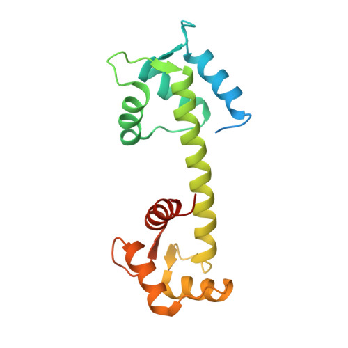
Crystal Structure of Wild-Type Centrin 1 from Mus musculus Occupied by Ca2.

Kim, S.Y.Kim, D.S.Hong, J.E.Park, J.H.

(2017) Biochemistry (Mosc) 82: 1129-1139

  • DOI: https://doi.org/10.1134/S0006297917100054
  • Primary Citation of Related Structures:  
    5D43

  • PubMed Abstract: 

    Mus musculus centrin 1 (MmCen1) is located at the cilium of photoreceptor cells connecting the outer segment through signal transduction components to the metabolically active inner segment. In the cilium, MmCen1 is involved in the translocation of transducin between compartments as a result of photoreceptor activation. In this study, we report the crystal structure of wild-type MmCen1 and its Ca2+-binding properties using structure-based mutagenesis. The crystal structure exhibits three structural features, i.e. four Ca2+ equally occupied at each EF-hand motif, structural changes accompanying helix motion at the N- and C-lobes, and adoption of N-C type dimerization when Ca2+ binds to EF-hand I and II in the N-lobe. The presence of MmCen1 dimers was confirmed in solution by native PAGE. Isothermal titration calorimetry data showed sequential binding of Ca2+ at four independent sites. Mutations S45A and D49A in EF-hand I alone disrupted the Ca2+-binding property of the wild-type protein. Based on the crystal structure of MmCen1, we suggest that a dimerization mode between the N- and C-lobes may be required by Ca2+ binding at the N-lobe.


  • Organizational Affiliation

    Chonbuk National University, College of Environmental & Bioresource Sciences, Division of Biotechnology, Iksan, 54596, Republic of Korea. junghee.park@jbnu.ac.kr.


Macromolecules
Find similar proteins by:  (by identity cutoff)  |  3D Structure
Entity ID: 1
MoleculeChains Sequence LengthOrganismDetailsImage
Centrin-1
A, B
178Mus musculusMutation(s): 0 
Gene Names: Cetn1Calt
UniProt
Find proteins for P41209 (Mus musculus)
Explore P41209 
Go to UniProtKB:  P41209
Entity Groups  
Sequence Clusters30% Identity50% Identity70% Identity90% Identity95% Identity100% Identity
UniProt GroupP41209
Sequence Annotations
Expand
  • Reference Sequence
Experimental Data & Validation

Experimental Data

  • Method: X-RAY DIFFRACTION
  • Resolution: 2.82 Å
  • R-Value Free: 0.329 
  • R-Value Work: 0.274 
  • R-Value Observed: 0.277 
  • Space Group: P 1 21 1
Unit Cell:
Length ( Å )Angle ( ˚ )
a = 49.75α = 90
b = 72.061β = 97.26
c = 62.248γ = 90
Software Package:
Software NamePurpose
REFMACrefinement
XDSdata processing
PHASERphasing
Cootmodel building
SCALAdata scaling

Structure Validation

View Full Validation Report


Entry History & Funding Information

Deposition Data


Funding OrganizationLocationGrant Number
National Research Foundation of KoreaKorea, Republic OfNRF-2012R1A1A1042354

Revision History  (Full details and data files)

  • Version 1.0: 2016-09-07
    Type: Initial release
  • Version 1.1: 2017-09-27
    Changes: Data collection, Derived calculations
  • Version 1.2: 2017-10-25
    Changes: Database references
  • Version 1.3: 2019-12-18
    Changes: Database references
  • Version 1.4: 2023-11-08
    Changes: Data collection, Database references, Derived calculations, Refinement description