5D0R

Crystal structure of human soluble Adenylyl Cyclase with the inhibitor bithionol


Experimental Data Snapshot

  • Method: X-RAY DIFFRACTION
  • Resolution: 2.24 Å
  • R-Value Free: 0.244 
  • R-Value Work: 0.177 
  • R-Value Observed: 0.181 

wwPDB Validation   3D Report Full Report


Ligand Structure Quality Assessment 


This is version 2.1 of the entry. See complete history


Literature

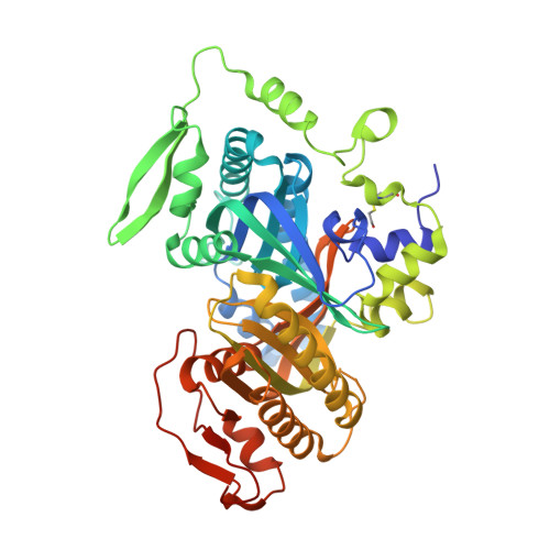
Bithionol Potently Inhibits Human Soluble Adenylyl Cyclase through Binding to the Allosteric Activator Site.

Kleinboelting, S.Ramos-Espiritu, L.Buck, H.Colis, L.van den Heuvel, J.Glickman, J.F.Levin, L.R.Buck, J.Steegborn, C.

(2016) J Biol Chem 291: 9776-9784

  • DOI: https://doi.org/10.1074/jbc.M115.708255
  • Primary Citation of Related Structures:  
    5D0R

  • PubMed Abstract: 

    The signaling molecule cAMP regulates functions ranging from bacterial transcription to mammalian memory. In mammals, cAMP is synthesized by nine transmembrane adenylyl cyclases (ACs) and one soluble AC (sAC). Despite similarities in their catalytic domains, these ACs differ in regulation. Transmembrane ACs respond to G proteins, whereas sAC is uniquely activated by bicarbonate. Via bicarbonate regulation, sAC acts as a physiological sensor for pH/bicarbonate/CO2, and it has been implicated as a therapeutic target, e.g. for diabetes, glaucoma, and a male contraceptive. Here we identify the bisphenols bithionol and hexachlorophene as potent, sAC-specific inhibitors. Inhibition appears mostly non-competitive with the substrate ATP, indicating that they act via an allosteric site. To analyze the interaction details, we solved a crystal structure of an sAC·bithionol complex. The structure reveals that the compounds are selective for sAC because they bind to the sAC-specific, allosteric binding site for the physiological activator bicarbonate. Structural comparison of the bithionol complex with apo-sAC and other sAC·ligand complexes along with mutagenesis experiments reveals an allosteric mechanism of inhibition; the compound induces rearrangements of substrate binding residues and of Arg(176), a trigger between the active site and allosteric site. Our results thus provide 1) novel insights into the communication between allosteric regulatory and active sites, 2) a novel mechanism for sAC inhibition, and 3) pharmacological compounds targeting this allosteric site and utilizing this mode of inhibition. These studies provide support for the future development of sAC-modulating drugs.


  • Organizational Affiliation

    From the Department of Biochemistry, University of Bayreuth, 95440 Bayreuth, Germany.


Macromolecules
Find similar proteins by:  (by identity cutoff)  |  3D Structure
Entity ID: 1
MoleculeChains Sequence LengthOrganismDetailsImage
Adenylate cyclase type 10475Homo sapiensMutation(s): 0 
Gene Names: ADCY10SAC
EC: 4.6.1.1
UniProt & NIH Common Fund Data Resources
Find proteins for Q96PN6 (Homo sapiens)
Explore Q96PN6 
Go to UniProtKB:  Q96PN6
PHAROS:  Q96PN6
GTEx:  ENSG00000143199 
Entity Groups  
Sequence Clusters30% Identity50% Identity70% Identity90% Identity95% Identity100% Identity
UniProt GroupQ96PN6
Sequence Annotations
Expand
  • Reference Sequence
Small Molecules
Ligands 5 Unique
IDChains Name / Formula / InChI Key2D Diagram3D Interactions
B1T
Query on B1T

Download Ideal Coordinates CCD File 
B [auth A]2,2'-sulfanediylbis(4,6-dichlorophenol)
C12 H6 Cl4 O2 S
JFIOVJDNOJYLKP-UHFFFAOYSA-N
GOL
Query on GOL

Download Ideal Coordinates CCD File 
M [auth A],
N [auth A]
GLYCEROL
C3 H8 O3
PEDCQBHIVMGVHV-UHFFFAOYSA-N
DMS
Query on DMS

Download Ideal Coordinates CCD File 
J [auth A],
K [auth A],
L [auth A]
DIMETHYL SULFOXIDE
C2 H6 O S
IAZDPXIOMUYVGZ-UHFFFAOYSA-N
EDO
Query on EDO

Download Ideal Coordinates CCD File 
D [auth A]
E [auth A]
F [auth A]
G [auth A]
H [auth A]
D [auth A],
E [auth A],
F [auth A],
G [auth A],
H [auth A],
I [auth A]
1,2-ETHANEDIOL
C2 H6 O2
LYCAIKOWRPUZTN-UHFFFAOYSA-N
ACT
Query on ACT

Download Ideal Coordinates CCD File 
C [auth A]ACETATE ION
C2 H3 O2
QTBSBXVTEAMEQO-UHFFFAOYSA-M
Modified Residues  1 Unique
IDChains TypeFormula2D DiagramParent
CME
Query on CME
A
L-PEPTIDE LINKINGC5 H11 N O3 S2CYS
Binding Affinity Annotations 
IDSourceBinding Affinity
B1T Binding MOAD:  5D0R Kd: 430 (nM) from 1 assay(s)
Experimental Data & Validation

Experimental Data

  • Method: X-RAY DIFFRACTION
  • Resolution: 2.24 Å
  • R-Value Free: 0.244 
  • R-Value Work: 0.177 
  • R-Value Observed: 0.181 
  • Space Group: P 63
Unit Cell:
Length ( Å )Angle ( ˚ )
a = 99.403α = 90
b = 99.403β = 90
c = 99.978γ = 120
Software Package:
Software NamePurpose
MOSFLMdata reduction
XSCALEdata scaling
MOLREPphasing
REFMACrefinement
PDB_EXTRACTdata extraction
Cootmodel building
XDSdata reduction

Structure Validation

View Full Validation Report


Ligand Structure Quality Assessment 


Entry History & Funding Information

Deposition Data


Funding OrganizationLocationGrant Number
German Research FoundationGermanySTE1701/11

Revision History  (Full details and data files)

  • Version 1.0: 2016-03-16
    Type: Initial release
  • Version 1.1: 2016-03-23
    Changes: Database references
  • Version 1.2: 2016-05-11
    Changes: Database references
  • Version 2.0: 2019-03-27
    Changes: Atomic model, Author supporting evidence, Data collection, Source and taxonomy
  • Version 2.1: 2024-01-10
    Changes: Data collection, Database references, Refinement description