


The crystal structure of mycobacterial Epoxide Hydrolase A
Schulz, E.C., Wilmanns, M., Henderson, S., Southall, S.To be published.
Experimental Data Snapshot
Entity ID: 1 | |||||
|---|---|---|---|---|---|
| Molecule | Chains | Sequence Length | Organism | Details | Image |
| Putative epoxide hydrolase EPHA | A [auth C], B [auth A], C [auth B], D | 325 | Mycolicibacterium thermoresistibile ATCC 19527 | Mutation(s): 0 Gene Names: KEK_07967 EC: 3.3.2.3 | 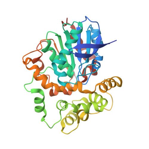 |
UniProt | |||||
Find proteins for G7CF24 (Mycolicibacterium thermoresistibile (strain ATCC 19527 / DSM 44167 / CIP 105390 / JCM 6362 / NCTC 10409 / 316)) Explore G7CF24 Go to UniProtKB: G7CF24 | |||||
Entity Groups | |||||
| Sequence Clusters | 30% Identity50% Identity70% Identity90% Identity95% Identity100% Identity | ||||
| UniProt Group | G7CF24 | ||||
Sequence AnnotationsExpand | |||||
| |||||
| Ligands 2 Unique | |||||
|---|---|---|---|---|---|
| ID | Chains | Name / Formula / InChI Key | 2D Diagram | 3D Interactions | |
| BSU Query on BSU | E [auth C], F [auth A], G [auth B], H [auth D] | 1,3-DIPHENYLUREA C13 H12 N2 O GWEHVDNNLFDJLR-UHFFFAOYSA-N |  | ||
| NA Query on NA | I [auth D] | SODIUM ION Na FKNQFGJONOIPTF-UHFFFAOYSA-N |  | ||
| Length ( Å ) | Angle ( ˚ ) |
|---|---|
| a = 61.8 | α = 90 |
| b = 105.01 | β = 90.11 |
| c = 108.95 | γ = 90 |
| Software Name | Purpose |
|---|---|
| PHENIX | refinement |
| XDS | data reduction |
| XSCALE | data scaling |
| PHASER | phasing |
RCSB PDB (citation) is hosted by
RCSB PDB is a member of the

