


Crystal structure of the first class E sortase transpeptidase, SrtE1 from Streptomyces coelicolor
Kattke, M.D., Cascio, D., Sawaya, M.R., Elliot, M.A., Clubb, R.T.To be published.
Experimental Data Snapshot
wwPDB Validation 3D Report Full Report

Entity ID: 1 | |||||
|---|---|---|---|---|---|
| Molecule | Chains | Sequence Length | Organism | Details | Image |
| SrtE1 | 195 | Streptomyces coelicolor A3(2) | Mutation(s): 0 Gene Names: SCO3850 | 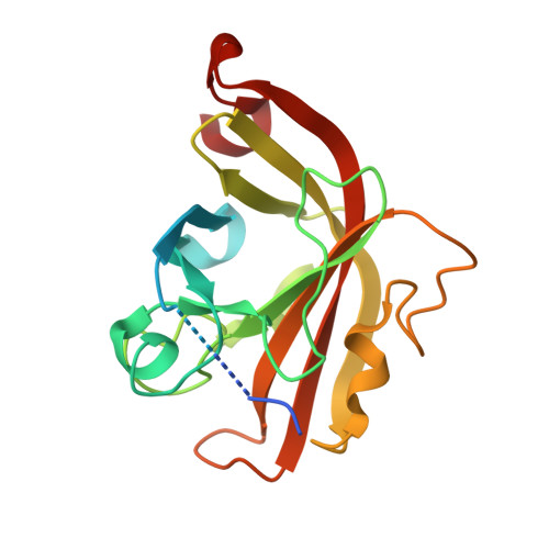 | |
UniProt | |||||
Find proteins for Q9XA14 (Streptomyces coelicolor (strain ATCC BAA-471 / A3(2) / M145)) Explore Q9XA14 Go to UniProtKB: Q9XA14 | |||||
Entity Groups | |||||
| Sequence Clusters | 30% Identity50% Identity70% Identity90% Identity95% Identity100% Identity | ||||
| UniProt Group | Q9XA14 | ||||
Sequence AnnotationsExpand | |||||
| |||||
| Ligands 1 Unique | |||||
|---|---|---|---|---|---|
| ID | Chains | Name / Formula / InChI Key | 2D Diagram | 3D Interactions | |
| GOL Query on GOL | B [auth A] | GLYCEROL C3 H8 O3 PEDCQBHIVMGVHV-UHFFFAOYSA-N |  | ||
| Length ( Å ) | Angle ( ˚ ) |
|---|---|
| a = 53.11 | α = 90 |
| b = 104.3 | β = 90 |
| c = 79.02 | γ = 90 |
| Software Name | Purpose |
|---|---|
| XSCALE | data scaling |
| PHASER | phasing |
| PHENIX | refinement |
| PDB_EXTRACT | data extraction |
| XDS | data reduction |
RCSB PDB (citation) is hosted by
RCSB PDB is a member of the

