5COB

Crystal structure of iridoid synthase in complex with NADP+ and 8-oxogeranial at 2.65-angstrom resolution


Experimental Data Snapshot

  • Method: X-RAY DIFFRACTION
  • Resolution: 2.65 Å
  • R-Value Free: 0.245 
  • R-Value Work: 0.194 
  • R-Value Observed: 0.197 

wwPDB Validation   3D Report Full Report


Ligand Structure Quality Assessment 


This is version 1.2 of the entry. See complete history


Literature

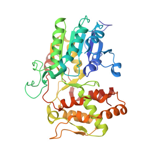
Structure of iridoid synthase in complex with NADP(+)/8-oxogeranial reveals the structural basis of its substrate specificity.

Qin, L.Zhu, Y.Ding, Z.Zhang, X.Ye, S.Zhang, R.

(2016) J Struct Biol 194: 224-230

  • DOI: https://doi.org/10.1016/j.jsb.2016.02.010
  • Primary Citation of Related Structures:  
    5COA, 5COB

  • PubMed Abstract: 

    Iridoid synthase (IS), as a vegetal enzyme belonging to the short-chain dehydrogenase/reductase (SDR) superfamily, produces the ring skeletons for downstream alkaloids with various pharmaceutical activities, including the commercially available antineoplastic agents, vinblastine and vincristine. Here, we present the crystal structures of IS in apo state and in complex with NADP(+)/8-oxogeranial, exhibiting an active center that lacks the classical Tyr/Lys/Ser triad spatially conserved in SDRs, with only the catalytically critical function of triad tyrosine remained in Tyr178. In consistent, mutation of Tyr178 to a phenylalanine residue significantly abolished the catalytic activity of IS. Within the substrate binding pocket, the linear-shaped 8-oxogeranial adopts an entirely extended conformation with its two aldehyde ends hydrogen-bonded to Tyr178-OH and Ser349-OH, respectively. In addition, the intermediate carbon chain of bound substrate is harbored by a well-ordered hydrophobic scaffold, involving residues Ile145, Phe149, Leu203, Met213, Phe342, Ile345 and Leu352. Mutagenesis studies showed that both Ser349 and the hydrophobic residues around are determinant to the substrate specificity and, consequently, the catalytic activity of IS. In contrast, the Gly150-Pro160 loop previously proposed as a factor involved in substrate binding might have very limited contribution, because the deletion of residues Ile151-His161 has only slight influence on the catalytic activity. We believe that the present work will help to elucidate the substrate specificity of IS and to integrate its detailed catalytic mechanism.


  • Organizational Affiliation

    National Laboratory of Biomacromolecules, Institute of Biophysics, Chinese Academy of Sciences, Beijing 100101, People's Republic of China; University of Chinese Academy of Sciences, Beijing 100049, People's Republic of China.


Macromolecules
Find similar proteins by:  (by identity cutoff)  |  3D Structure
Entity ID: 1
MoleculeChains Sequence LengthOrganismDetailsImage
Iridoid synthase
A, B, C, D
365Catharanthus roseusMutation(s): 0 
EC: 1.3.1.99
UniProt
Find proteins for K7WDL7 (Catharanthus roseus)
Explore K7WDL7 
Go to UniProtKB:  K7WDL7
Entity Groups  
Sequence Clusters30% Identity50% Identity70% Identity90% Identity95% Identity100% Identity
UniProt GroupK7WDL7
Sequence Annotations
Expand
  • Reference Sequence
Small Molecules
Ligands 3 Unique
IDChains Name / Formula / InChI Key2D Diagram3D Interactions
NAP
Query on NAP

Download Ideal Coordinates CCD File 
E [auth A],
H [auth B],
K [auth C],
N [auth D]
NADP NICOTINAMIDE-ADENINE-DINUCLEOTIDE PHOSPHATE
C21 H28 N7 O17 P3
XJLXINKUBYWONI-NNYOXOHSSA-N
PG4
Query on PG4

Download Ideal Coordinates CCD File 
G [auth A],
J [auth B],
M [auth C],
P [auth D]
TETRAETHYLENE GLYCOL
C8 H18 O5
UWHCKJMYHZGTIT-UHFFFAOYSA-N
XOG
Query on XOG

Download Ideal Coordinates CCD File 
F [auth A],
I [auth B],
L [auth C],
O [auth D]
(2E,6E)-2,6-dimethylocta-2,6-dienedial
C10 H14 O2
GRHWFPUCRVCMRY-TXFIJWAUSA-N
Experimental Data & Validation

Experimental Data

  • Method: X-RAY DIFFRACTION
  • Resolution: 2.65 Å
  • R-Value Free: 0.245 
  • R-Value Work: 0.194 
  • R-Value Observed: 0.197 
  • Space Group: P 1 21 1
Unit Cell:
Length ( Å )Angle ( ˚ )
a = 65.988α = 90
b = 171.914β = 89.72
c = 66.032γ = 90
Software Package:
Software NamePurpose
PHENIXrefinement
HKL-2000data reduction
HKL-2000data scaling
PHASERphasing

Structure Validation

View Full Validation Report


Ligand Structure Quality Assessment 


Entry History 

Deposition Data

Revision History  (Full details and data files)

  • Version 1.0: 2016-03-09
    Type: Initial release
  • Version 1.1: 2016-03-30
    Changes: Database references
  • Version 1.2: 2024-03-20
    Changes: Data collection, Database references, Derived calculations