5CO8

Crystal structure of the Holliday junction-resolving enzyme GEN1 (WT) in complex with product DNA and Mg2+ ion


Experimental Data Snapshot

  • Method: X-RAY DIFFRACTION
  • Resolution: 2.40 Å
  • R-Value Free: 0.255 
  • R-Value Work: 0.239 
  • R-Value Observed: 0.240 

wwPDB Validation   3D Report Full Report


This is version 1.2 of the entry. See complete history


Literature

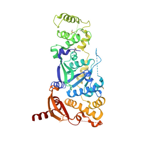
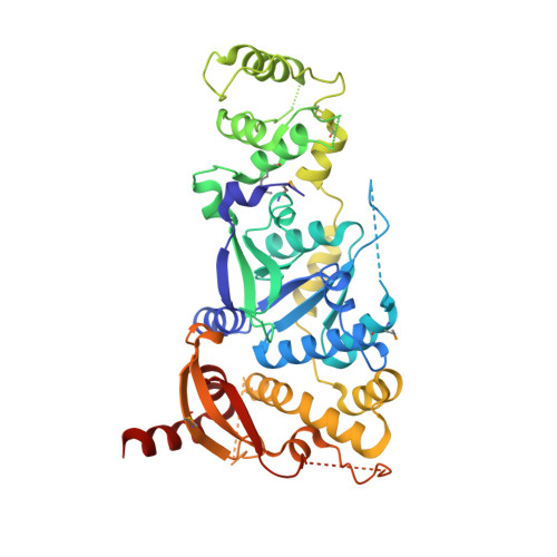
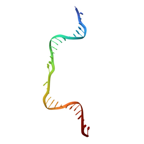
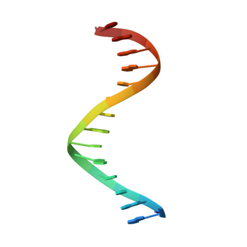
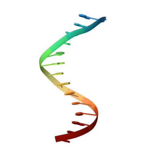
Crystal Structure of a Eukaryotic GEN1 Resolving Enzyme Bound to DNA.

Liu, Y.Freeman, A.D.Declais, A.C.Wilson, T.J.Gartner, A.Lilley, D.M.

(2015) Cell Rep 13: 2565-2575

  • DOI: https://doi.org/10.1016/j.celrep.2015.11.042
  • Primary Citation of Related Structures:  
    5CNQ, 5CO8

  • PubMed Abstract: 

    We present the crystal structure of the junction-resolving enzyme GEN1 bound to DNA at 2.5 Å resolution. The structure of the GEN1 protein reveals it to have an elaborated FEN-XPG family fold that is modified for its role in four-way junction resolution. The functional unit in the crystal is a monomer of active GEN1 bound to the product of resolution cleavage, with an extensive DNA binding interface for both helical arms. Within the crystal lattice, a GEN1 dimer interface juxtaposes two products, whereby they can be reconnected into a four-way junction, the structure of which agrees with that determined in solution. The reconnection requires some opening of the DNA structure at the center, in agreement with permanganate probing and 2-aminopurine fluorescence. The structure shows that a relaxation of the DNA structure accompanies cleavage, suggesting how second-strand cleavage is accelerated to ensure productive resolution of the junction.


  • Organizational Affiliation

    Cancer Research UK Nucleic Acid Structure Research Group, MSI/WTB Complex, University of Dundee, Dow Street, Dundee DD1 5EH, UK.


Macromolecules

Find similar proteins by:  (by identity cutoff)  |  3D Structure
Entity ID: 2
MoleculeChains Sequence LengthOrganismDetailsImage
Nuclease-like proteinB [auth C]464Thermochaetoides thermophila DSM 1495Mutation(s): 0 
Gene Names: CTHT_0007290
UniProt
Find proteins for G0RYN2 (Chaetomium thermophilum (strain DSM 1495 / CBS 144.50 / IMI 039719))
Explore G0RYN2 
Go to UniProtKB:  G0RYN2
Entity Groups  
Sequence Clusters30% Identity50% Identity70% Identity90% Identity95% Identity100% Identity
UniProt GroupG0RYN2
Sequence Annotations
Expand
  • Reference Sequence
Find similar proteins by:  (by identity cutoff)  |  3D Structure
Entity ID: 4
MoleculeChains Sequence LengthOrganismDetailsImage
Nuclease-like proteinD [auth A]464Thermochaetoides thermophila DSM 1495Mutation(s): 0 
Gene Names: CTHT_0007290
UniProt
Find proteins for G0RYN2 (Chaetomium thermophilum (strain DSM 1495 / CBS 144.50 / IMI 039719))
Explore G0RYN2 
Go to UniProtKB:  G0RYN2
Entity Groups  
Sequence Clusters30% Identity50% Identity70% Identity90% Identity95% Identity100% Identity
UniProt GroupG0RYN2
Sequence Annotations
Expand
  • Reference Sequence
Find similar nucleic acids by:  (by identity cutoff)  |  3D Structure
Entity ID: 1
MoleculeChains LengthOrganismImage
DNA (31-MER)A [auth R]31synthetic construct
Sequence Annotations
Expand
  • Reference Sequence

Find similar nucleic acids by:  Sequence   |   3D Structure  

Entity ID: 3
MoleculeChains LengthOrganismImage
DNA (5'-D(*AP*GP*AP*CP*TP*GP*CP*AP*GP*TP*TP*GP*AP*GP*TP*C)-3')C [auth X]16synthetic construct
Sequence Annotations
Expand
  • Reference Sequence

Find similar nucleic acids by:  Sequence   |   3D Structure  

Entity ID: 5
MoleculeChains LengthOrganismImage
DNA (5'-D(*TP*GP*AP*GP*CP*GP*GP*TP*GP*GP*TP*TP*GP*GP*A)-3')E [auth H]15synthetic construct
Sequence Annotations
Expand
  • Reference Sequence
Small Molecules
Modified Residues  1 Unique
IDChains TypeFormula2D DiagramParent
MSE
Query on MSE
B [auth C]L-PEPTIDE LINKINGC5 H11 N O2 SeMET
Experimental Data & Validation

Experimental Data

  • Method: X-RAY DIFFRACTION
  • Resolution: 2.40 Å
  • R-Value Free: 0.255 
  • R-Value Work: 0.239 
  • R-Value Observed: 0.240 
  • Space Group: P 31 2 1
Unit Cell:
Length ( Å )Angle ( ˚ )
a = 98.072α = 90
b = 98.072β = 90
c = 119.571γ = 120
Software Package:
Software NamePurpose
PHENIXrefinement
SCALAdata scaling
PDB_EXTRACTdata extraction
PHENIXphasing

Structure Validation

View Full Validation Report


Entry History & Funding Information

Deposition Data


Funding OrganizationLocationGrant Number
Cancer Research UKUnited Kingdom--

Revision History  (Full details and data files)

  • Version 1.0: 2016-01-13
    Type: Initial release
  • Version 1.1: 2018-10-24
    Changes: Advisory, Data collection, Derived calculations
  • Version 1.2: 2018-11-21
    Changes: Data collection, Derived calculations