5CI7

Structure of ULK1 bound to a selective inhibitor


Experimental Data Snapshot

  • Method: X-RAY DIFFRACTION
  • Resolution: 1.74 Å
  • R-Value Free: 0.207 
  • R-Value Work: 0.169 
  • R-Value Observed: 0.171 

wwPDB Validation   3D Report Full Report


Ligand Structure Quality Assessment 


This is version 1.4 of the entry. See complete history


Literature

Discovery and structure of a new inhibitor scaffold of the autophagy initiating kinase ULK1.

Lazarus, M.B.Shokat, K.M.

(2015) Bioorg Med Chem 23: 5483-5488

  • DOI: https://doi.org/10.1016/j.bmc.2015.07.034
  • Primary Citation of Related Structures:  
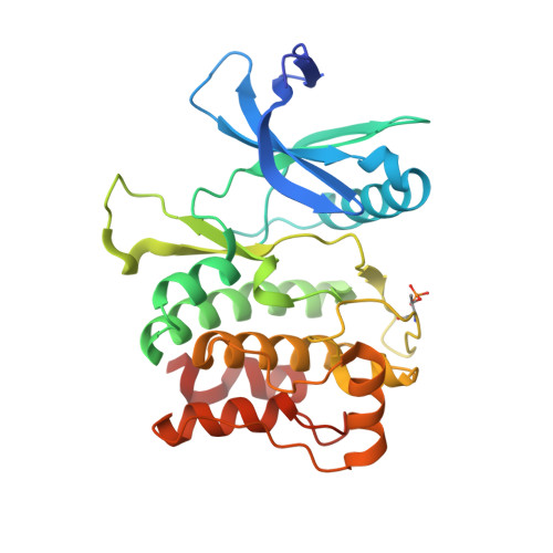
    5CI7

  • PubMed Abstract: 

    Energy homeostasis in eukaryotic cells is a complex and fundamental process that is misregulated in several human diseases. A key component of energy regulation is a process called autophagy that involves the recycling of cellular components. There has been much recent interest in studying the mechanism of autophagy to understand an important cellular process and to evaluate the therapeutic potential in targeting autophagy. Activation of a kinase called ULK1 initiates autophagy by driving downstream pathways that lead to the formation of double membrane bound vesicles that surround the cellular contents that are to be degraded. Here, we report the discovery of an inhibitor of ULK1 with improved selectivity and a high-resolution crystal structure of the compound bound to the kinase, which will be useful tools for studying autophagy in cells.


  • Organizational Affiliation

    Howard Hughes Medical Institute and Department of Cellular and Molecular Pharmacology, University of California, San Francisco, San Francisco, CA 94158, USA.


Macromolecules
Find similar proteins by:  (by identity cutoff)  |  3D Structure
Entity ID: 1
MoleculeChains Sequence LengthOrganismDetailsImage
Serine/threonine-protein kinase ULK1287Homo sapiensMutation(s): 2 
Gene Names: ULK1KIAA0722
EC: 2.7.11.1
UniProt & NIH Common Fund Data Resources
Find proteins for O75385 (Homo sapiens)
Explore O75385 
Go to UniProtKB:  O75385
PHAROS:  O75385
GTEx:  ENSG00000177169 
Entity Groups  
Sequence Clusters30% Identity50% Identity70% Identity90% Identity95% Identity100% Identity
UniProt GroupO75385
Sequence Annotations
Expand
  • Reference Sequence
Small Molecules
Modified Residues  1 Unique
IDChains TypeFormula2D DiagramParent
TPO
Query on TPO
A
L-PEPTIDE LINKINGC4 H10 N O6 PTHR
Binding Affinity Annotations 
IDSourceBinding Affinity
51W Binding MOAD:  5CI7 IC50: 120 (nM) from 1 assay(s)
BindingDB:  5CI7 IC50: 120 (nM) from 1 assay(s)
Experimental Data & Validation

Experimental Data

  • Method: X-RAY DIFFRACTION
  • Resolution: 1.74 Å
  • R-Value Free: 0.207 
  • R-Value Work: 0.169 
  • R-Value Observed: 0.171 
  • Space Group: P 42 21 2
Unit Cell:
Length ( Å )Angle ( ˚ )
a = 66.86α = 90
b = 66.86β = 90
c = 116.61γ = 90
Software Package:
Software NamePurpose
PHENIXrefinement
iMOSFLMdata reduction
SCALAdata scaling
PHASERphasing

Structure Validation

View Full Validation Report


Ligand Structure Quality Assessment 


Entry History & Funding Information

Deposition Data


Funding OrganizationLocationGrant Number
National Institutes of Health/National Institute Of Allergy and Infectious Diseases (NIH/NIAID)United StatesR01AI099245

Revision History  (Full details and data files)

  • Version 1.0: 2015-08-26
    Type: Initial release
  • Version 1.1: 2015-09-09
    Changes: Database references
  • Version 1.2: 2017-09-27
    Changes: Author supporting evidence, Database references, Derived calculations
  • Version 1.3: 2019-12-11
    Changes: Author supporting evidence
  • Version 1.4: 2023-09-27
    Changes: Data collection, Database references, Refinement description