5CHT

Crystal structure of USP18


Experimental Data Snapshot

  • Method: X-RAY DIFFRACTION
  • Resolution: 2.80 Å
  • R-Value Free: 0.283 
  • R-Value Work: 0.225 
  • R-Value Observed: 0.228 

wwPDB Validation   3D Report Full Report


This is version 1.3 of the entry. See complete history


Literature

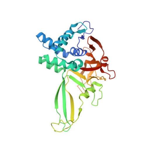
Structural basis of the specificity of USP18 toward ISG15.

Basters, A.Geurink, P.P.Rocker, A.Witting, K.F.Tadayon, R.Hess, S.Semrau, M.S.Storici, P.Ovaa, H.Knobeloch, K.P.Fritz, G.

(2017) Nat Struct Mol Biol 24: 270-278

  • DOI: https://doi.org/10.1038/nsmb.3371
  • Primary Citation of Related Structures:  
    5CHT, 5CHV

  • PubMed Abstract: 

    Protein modification by ubiquitin and ubiquitin-like modifiers (Ubls) is counteracted by ubiquitin proteases and Ubl proteases, collectively termed DUBs. In contrast to other proteases of the ubiquitin-specific protease (USP) family, USP18 shows no reactivity toward ubiquitin but specifically deconjugates the interferon-induced Ubl ISG15. To identify the molecular determinants of this specificity, we solved the crystal structures of mouse USP18 alone and in complex with mouse ISG15. USP18 was crystallized in an open and a closed conformation, thus revealing high flexibility of the enzyme. Structural data, biochemical and mutational analysis showed that only the C-terminal ubiquitin-like domain of ISG15 is recognized and essential for USP18 activity. A critical hydrophobic patch in USP18 interacts with a hydrophobic region unique to ISG15, thus providing evidence that USP18's ISG15 specificity is mediated by a small interaction interface. Our results may provide a structural basis for the development of new drugs modulating ISG15 linkage.


  • Organizational Affiliation

    Institute of Neuropathology, Faculty of Medicine, University of Freiburg, Freiburg, Germany, Breisacherstraße 64, 79106 Freiburg, Germany.


Macromolecules
Find similar proteins by:  (by identity cutoff)  |  3D Structure
Entity ID: 1
MoleculeChains Sequence LengthOrganismDetailsImage
Ubl carboxyl-terminal hydrolase 18
A, B
326Mus musculusMutation(s): 0 
Gene Names: Usp18Ubp43
EC: 3.4.19
UniProt
Find proteins for Q9WTV6 (Mus musculus)
Explore Q9WTV6 
Go to UniProtKB:  Q9WTV6
Entity Groups  
Sequence Clusters30% Identity50% Identity70% Identity90% Identity95% Identity100% Identity
UniProt GroupQ9WTV6
Sequence Annotations
Expand
  • Reference Sequence
Experimental Data & Validation

Experimental Data

  • Method: X-RAY DIFFRACTION
  • Resolution: 2.80 Å
  • R-Value Free: 0.283 
  • R-Value Work: 0.225 
  • R-Value Observed: 0.228 
  • Space Group: P 21 21 21
Unit Cell:
Length ( Å )Angle ( ˚ )
a = 53.959α = 90
b = 89.747β = 90
c = 149.406γ = 90
Software Package:
Software NamePurpose
PHENIXrefinement
XDSdata reduction
XSCALEdata scaling
PHASERphasing
Cootmodel building

Structure Validation

View Full Validation Report


Entry History 

Deposition Data

Revision History  (Full details and data files)

  • Version 1.0: 2016-06-29
    Type: Initial release
  • Version 1.1: 2017-02-15
    Changes: Database references
  • Version 1.2: 2017-03-15
    Changes: Database references
  • Version 1.3: 2024-01-10
    Changes: Data collection, Database references, Derived calculations, Refinement description