5CFC

Crystal Structure of Human Cardiovirus SAFV-3


Experimental Data Snapshot

  • Method: X-RAY DIFFRACTION
  • Resolution: 2.50 Å
  • R-Value Work: 0.220 

wwPDB Validation   3D Report Full Report


This is version 1.3 of the entry. See complete history


Literature

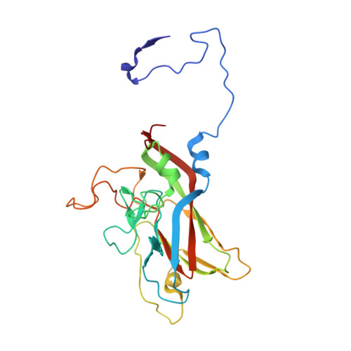
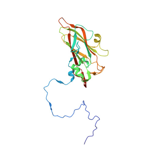
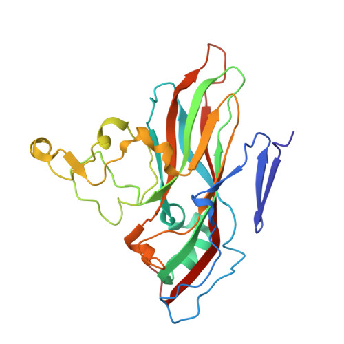
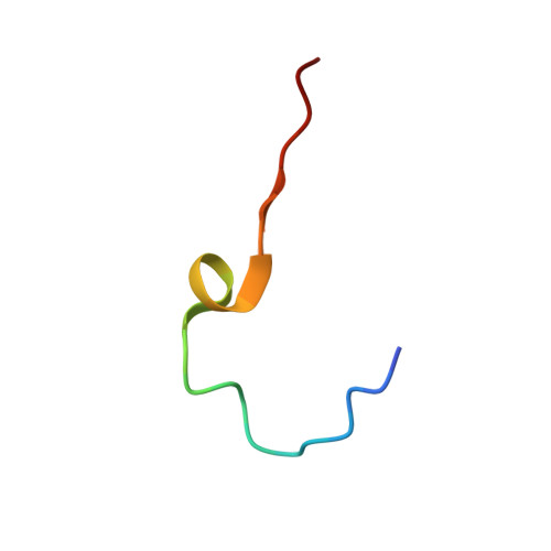
Structure and Genome Release Mechanism of the Human Cardiovirus Saffold Virus 3.

Mullapudi, E.Novacek, J.Palkova, L.Kulich, P.Lindberg, A.M.van Kuppeveld, F.J.Plevka, P.

(2016) J Virol 90: 7628-7639

  • DOI: https://doi.org/10.1128/JVI.00746-16
  • Primary Citation of Related Structures:  
    5A8F, 5CFC, 5CFD

  • PubMed Abstract: 

    In order to initiate an infection, viruses need to deliver their genomes into cells. This involves uncoating the genome and transporting it to the cytoplasm. The process of genome delivery is not well understood for nonenveloped viruses. We address this gap in our current knowledge by studying the uncoating of the nonenveloped human cardiovirus Saffold virus 3 (SAFV-3) of the family Picornaviridae SAFVs cause diseases ranging from gastrointestinal disorders to meningitis. We present a structure of a native SAFV-3 virion determined to 2.5 Å by X-ray crystallography and an 11-Å-resolution cryo-electron microscopy reconstruction of an "altered" particle that is primed for genome release. The altered particles are expanded relative to the native virus and contain pores in the capsid that might serve as channels for the release of VP4 subunits, N termini of VP1, and the RNA genome. Unlike in the related enteroviruses, pores in SAFV-3 are located roughly between the icosahedral 3- and 5-fold axes at an interface formed by two VP1 and one VP3 subunit. Furthermore, in native conditions many cardioviruses contain a disulfide bond formed by cysteines that are separated by just one residue. The disulfide bond is located in a surface loop of VP3. We determined the structure of the SAFV-3 virion in which the disulfide bonds are reduced. Disruption of the bond had minimal effect on the structure of the loop, but it increased the stability and decreased the infectivity of the virus. Therefore, compounds specifically disrupting or binding to the disulfide bond might limit SAFV infection.


  • Organizational Affiliation

    Central European Institute of Technology, Masaryk University, Brno, Czech Republic.


Macromolecules
Find similar proteins by:  (by identity cutoff)  |  3D Structure
Entity ID: 1
MoleculeChains Sequence LengthOrganismDetailsImage
VP1252Saffold virusMutation(s): 0 
UniProt
Find proteins for C0MHL9 (Saffold virus)
Explore C0MHL9 
Go to UniProtKB:  C0MHL9
Entity Groups  
Sequence Clusters30% Identity50% Identity70% Identity90% Identity95% Identity100% Identity
UniProt GroupC0MHL9
Sequence Annotations
Expand
  • Reference Sequence
Find similar proteins by:  (by identity cutoff)  |  3D Structure
Entity ID: 2
MoleculeChains Sequence LengthOrganismDetailsImage
VP3232Saffold virusMutation(s): 0 
UniProt
Find proteins for E3TMG8 (Saffold virus)
Explore E3TMG8 
Go to UniProtKB:  E3TMG8
Entity Groups  
Sequence Clusters30% Identity50% Identity70% Identity90% Identity95% Identity100% Identity
UniProt GroupE3TMG8
Sequence Annotations
Expand
  • Reference Sequence
Find similar proteins by:  (by identity cutoff)  |  3D Structure
Entity ID: 3
MoleculeChains Sequence LengthOrganismDetailsImage
VP2258Saffold virusMutation(s): 0 
UniProt
Find proteins for C0MHL9 (Saffold virus)
Explore C0MHL9 
Go to UniProtKB:  C0MHL9
Entity Groups  
Sequence Clusters30% Identity50% Identity70% Identity90% Identity95% Identity100% Identity
UniProt GroupC0MHL9
Sequence Annotations
Expand
  • Reference Sequence

Find similar proteins by:  Sequence   |   3D Structure  

Entity ID: 4
MoleculeChains Sequence LengthOrganismDetailsImage
VP424Saffold virusMutation(s): 0 
UniProt
Find proteins for C3U5A3 (Saffold virus 5)
Explore C3U5A3 
Go to UniProtKB:  C3U5A3
Entity Groups  
Sequence Clusters30% Identity50% Identity70% Identity90% Identity95% Identity100% Identity
UniProt GroupC3U5A3
Sequence Annotations
Expand
  • Reference Sequence
Experimental Data & Validation

Experimental Data

  • Method: X-RAY DIFFRACTION
  • Resolution: 2.50 Å
  • R-Value Work: 0.220 
  • Space Group: P 32 2 1
Unit Cell:
Length ( Å )Angle ( ˚ )
a = 300.502α = 90
b = 300.502β = 90
c = 722.103γ = 120
Software Package:
Software NamePurpose
XDSdata reduction
PHASERphasing
CNSrefinement
PDB_EXTRACTdata extraction
CNSphasing

Structure Validation

View Full Validation Report


Entry History 

Deposition Data

Revision History  (Full details and data files)

  • Version 1.0: 2016-06-15
    Type: Initial release
  • Version 1.1: 2016-06-22
    Changes: Database references
  • Version 1.2: 2016-08-24
    Changes: Database references
  • Version 1.3: 2018-04-11
    Changes: Data collection, Source and taxonomy, Structure summary