5CE9

structure of tyrosinase from walnut (Juglans regia)


Experimental Data Snapshot

  • Method: X-RAY DIFFRACTION
  • Resolution: 1.80 Å
  • R-Value Free: 0.188 
  • R-Value Work: 0.170 
  • R-Value Observed: 0.171 

wwPDB Validation   3D Report Full Report


This is version 1.5 of the entry. See complete history


Literature

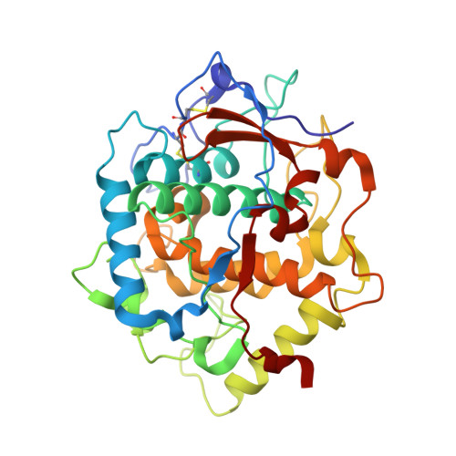
The Structure of a Plant Tyrosinase from Walnut Leaves Reveals the Importance of """"Substrate-Guiding Residues"""" for Enzymatic Specificity.

Bijelic, A.Pretzler, M.Molitor, C.Zekiri, F.Rompel, A.

(2015) Angew Chem Int Ed Engl 54: 14677-14680

  • DOI: https://doi.org/10.1002/anie.201506994
  • Primary Citation of Related Structures:  
    5CE9

  • PubMed Abstract: 

    Tyrosinases and catechol oxidases are members of the class of type III copper enzymes. While tyrosinases accept both mono- and o-diphenols as substrates, only the latter substrate is converted by catechol oxidases. Researchers have been working for decades to elucidate the monophenolase/diphenolase specificity on a structural level and have introduced an early hypothesis that states that the reason for the lack of monophenolase activity in catechol oxidases may be its structurally restricted active site. However, recent structural and biochemical studies of this enzyme class have raised doubts about this theory. Herein, the first crystal structure of a plant tyrosinase (from Juglans regia) is presented. The structure reveals that the distinction between mono- and diphenolase activity does not depend on the degree of restriction of the active site, and thus a more important role for amino acid residues located at the entrance to and in the second shell of the active site is proposed.


  • Organizational Affiliation

    Institut für Biophysikalische Chemie, Fakultät für Chemie, Universität Wien, Althanstraße 14, 1090 Wien (Austria) http://www.bpc.univie.ac.at.


Macromolecules
Find similar proteins by:  (by identity cutoff)  |  3D Structure
Entity ID: 1
MoleculeChains Sequence LengthOrganismDetailsImage
Polyphenol oxidase
A, B
339Juglans regiaMutation(s): 0 
UniProt
Find proteins for C0LU17 (Juglans regia)
Explore C0LU17 
Go to UniProtKB:  C0LU17
Entity Groups  
Sequence Clusters30% Identity50% Identity70% Identity90% Identity95% Identity100% Identity
UniProt GroupC0LU17
Sequence Annotations
Expand
  • Reference Sequence
Small Molecules
Ligands 3 Unique
IDChains Name / Formula / InChI Key2D Diagram3D Interactions
CU
Query on CU

Download Ideal Coordinates CCD File 
C [auth A],
D [auth A],
I [auth B],
J [auth B]
COPPER (II) ION
Cu
JPVYNHNXODAKFH-UHFFFAOYSA-N
NA
Query on NA

Download Ideal Coordinates CCD File 
F [auth A]
G [auth A]
H [auth A]
L [auth B]
M [auth B]
F [auth A],
G [auth A],
H [auth A],
L [auth B],
M [auth B],
N [auth B],
O [auth B]
SODIUM ION
Na
FKNQFGJONOIPTF-UHFFFAOYSA-N
O
Query on O

Download Ideal Coordinates CCD File 
E [auth A],
K [auth B]
OXYGEN ATOM
O
XLYOFNOQVPJJNP-UHFFFAOYSA-N
Experimental Data & Validation

Experimental Data

  • Method: X-RAY DIFFRACTION
  • Resolution: 1.80 Å
  • R-Value Free: 0.188 
  • R-Value Work: 0.170 
  • R-Value Observed: 0.171 
  • Space Group: C 1 2 1
Unit Cell:
Length ( Å )Angle ( ˚ )
a = 115.43α = 90
b = 90.94β = 130.18
c = 86.58γ = 90
Software Package:
Software NamePurpose
PHENIXrefinement
XDSdata reduction
PHASERphasing

Structure Validation

View Full Validation Report


Entry History & Funding Information

Deposition Data


Funding OrganizationLocationGrant Number
Austrian Science FundAustriaP25217-N28

Revision History  (Full details and data files)

  • Version 1.0: 2015-10-28
    Type: Initial release
  • Version 1.1: 2016-01-13
    Changes: Database references
  • Version 1.2: 2016-03-30
    Changes: Data collection
  • Version 1.3: 2017-09-06
    Changes: Author supporting evidence
  • Version 1.4: 2019-02-20
    Changes: Advisory, Data collection, Derived calculations
  • Version 1.5: 2024-01-10
    Changes: Data collection, Database references, Derived calculations, Refinement description