5CDC

Crystal Structure of Israel acute Paralysis Virus


Experimental Data Snapshot

  • Method: X-RAY DIFFRACTION
  • Resolution: 4.00 Å

wwPDB Validation   3D Report Full Report


This is version 1.5 of the entry. See complete history


Literature

Virion Structure of Iflavirus Slow Bee Paralysis Virus at 2.6-Angstrom Resolution.

Kalynych, S.Pridal, A.Palkova, L.Levdansky, Y.de Miranda, J.R.Plevka, P.

(2016) J Virol 90: 7444-7455

  • DOI: https://doi.org/10.1128/JVI.00680-16
  • Primary Citation of Related Structures:  
    5CDC, 5CDD, 5J96, 5J98

  • PubMed Abstract: 

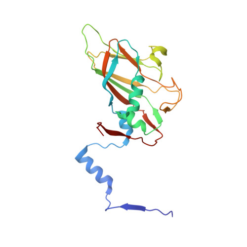
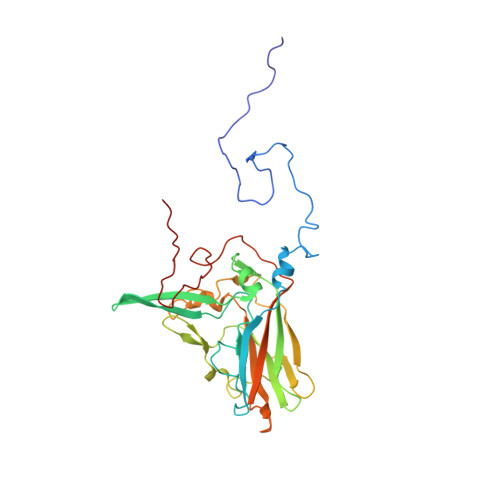
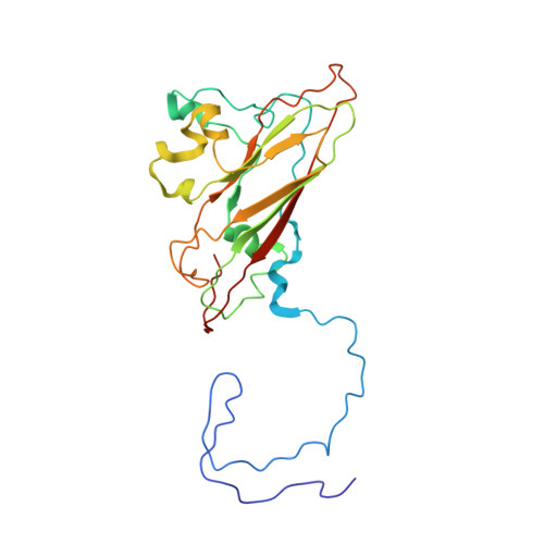
    The western honeybee (Apis mellifera) is the most important commercial insect pollinator. However, bees are under pressure from habitat loss, environmental stress, and pathogens, including viruses that can cause lethal epidemics. Slow bee paralysis virus (SBPV) belongs to the Iflaviridae family of nonenveloped single-stranded RNA viruses. Here we present the structure of the SBPV virion determined from two crystal forms to resolutions of 3.4 Å and 2.6 Å. The overall structure of the virion resembles that of picornaviruses, with the three major capsid proteins VP1 to 3 organized into a pseudo-T3 icosahedral capsid. However, the SBPV capsid protein VP3 contains a C-terminal globular domain that has not been observed in other viruses from the order Picornavirales The protruding (P) domains form "crowns" on the virion surface around each 5-fold axis in one of the crystal forms. However, the P domains are shifted 36 Å toward the 3-fold axis in the other crystal form. Furthermore, the P domain contains the Ser-His-Asp triad within a surface patch of eight conserved residues that constitutes a putative catalytic or receptor-binding site. The movements of the domain might be required for efficient substrate cleavage or receptor binding during virus cell entry. In addition, capsid protein VP2 contains an RGD sequence that is exposed on the virion surface, indicating that integrins might be cellular receptors of SBPV.


  • Organizational Affiliation

    Structural Virology, Central European Institute of Technology, Masaryk University, Brno, Czech Republic.


Macromolecules
Find similar proteins by:  (by identity cutoff)  |  3D Structure
Entity ID: 1
MoleculeChains Sequence LengthOrganismDetailsImage
VP1, Structural polyprotein203Israeli acute paralysis virusMutation(s): 0 
UniProt
Find proteins for D1FK67 (Israeli acute paralysis virus)
Explore D1FK67 
Go to UniProtKB:  D1FK67
Entity Groups  
Sequence Clusters30% Identity50% Identity70% Identity90% Identity95% Identity100% Identity
UniProt GroupD1FK67
Sequence Annotations
Expand
  • Reference Sequence
Find similar proteins by:  (by identity cutoff)  |  3D Structure
Entity ID: 2
MoleculeChains Sequence LengthOrganismDetailsImage
VP2, Structural polyprotein300Israeli acute paralysis virusMutation(s): 0 
UniProt
Find proteins for D1FK67 (Israeli acute paralysis virus)
Explore D1FK67 
Go to UniProtKB:  D1FK67
Entity Groups  
Sequence Clusters30% Identity50% Identity70% Identity90% Identity95% Identity100% Identity
UniProt GroupD1FK67
Sequence Annotations
Expand
  • Reference Sequence
Find similar proteins by:  (by identity cutoff)  |  3D Structure
Entity ID: 3
MoleculeChains Sequence LengthOrganismDetailsImage
VP3, Structural polyprotein243Israeli acute paralysis virusMutation(s): 0 
UniProt
Find proteins for D1FK67 (Israeli acute paralysis virus)
Explore D1FK67 
Go to UniProtKB:  D1FK67
Entity Groups  
Sequence Clusters30% Identity50% Identity70% Identity90% Identity95% Identity100% Identity
UniProt GroupD1FK67
Sequence Annotations
Expand
  • Reference Sequence
Find similar proteins by:  (by identity cutoff)  |  3D Structure
Entity ID: 4
MoleculeChains Sequence LengthOrganismDetailsImage
VP445Israeli acute paralysis virusMutation(s): 0 
Sequence Annotations
Expand
  • Reference Sequence
Experimental Data & Validation

Experimental Data

  • Method: X-RAY DIFFRACTION
  • Resolution: 4.00 Å
  • Space Group: P 21 21 2
Unit Cell:
Length ( Å )Angle ( ˚ )
a = 343.149α = 90
b = 383.261β = 90
c = 329.911γ = 90
Software Package:
Software NamePurpose
CNSrefinement
PHASERphasing
PDB_EXTRACTdata extraction
Aimlessdata scaling
PHASERphasing

Structure Validation

View Full Validation Report


Entry History 

Deposition Data

Revision History  (Full details and data files)

  • Version 1.0: 2016-07-06
    Type: Initial release
  • Version 1.1: 2016-07-20
    Changes: Database references
  • Version 1.2: 2016-08-24
    Changes: Database references
  • Version 1.3: 2018-04-11
    Changes: Data collection
  • Version 1.4: 2018-05-30
    Changes: Data collection, Source and taxonomy
  • Version 1.5: 2024-01-10
    Changes: Data collection, Database references, Derived calculations, Refinement description