5CCV

Crystal structure of full-length NS5 from dengue virus type 3


Experimental Data Snapshot

  • Method: X-RAY DIFFRACTION
  • Resolution: 3.60 Å
  • R-Value Free: 0.274 
  • R-Value Work: 0.238 
  • R-Value Observed: 0.239 

wwPDB Validation   3D Report Full Report


Ligand Structure Quality Assessment 


This is version 1.4 of the entry. See complete history


Literature

Dengue Virus Nonstructural Protein 5 (NS5) Assembles into a Dimer with a Unique Methyltransferase and Polymerase Interface.

Klema, V.J.Ye, M.Hindupur, A.Teramoto, T.Gottipati, K.Padmanabhan, R.Choi, K.H.

(2016) PLoS Pathog 12: e1005451-e1005451

  • DOI: https://doi.org/10.1371/journal.ppat.1005451
  • Primary Citation of Related Structures:  
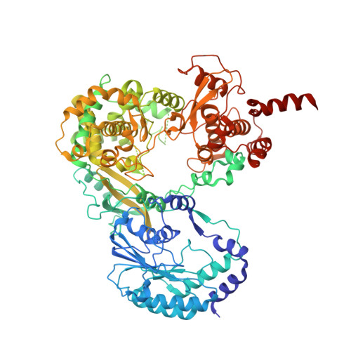
    5CCV

  • PubMed Abstract: 

    Flavivirus nonstructural protein 5 (NS5) consists of methyltransferase (MTase) and RNA-dependent RNA polymerase (RdRp) domains, which catalyze 5'-RNA capping/methylation and RNA synthesis, respectively, during viral genome replication. Although the crystal structure of flavivirus NS5 is known, no data about the quaternary organization of the functional enzyme are available. We report the crystal structure of dengue virus full-length NS5, where eight molecules of NS5 are arranged as four independent dimers in the crystallographic asymmetric unit. The relative orientation of each monomer within the dimer, as well as the orientations of the MTase and RdRp domains within each monomer, is conserved, suggesting that these structural arrangements represent the biologically relevant conformation and assembly of this multi-functional enzyme. Essential interactions between MTase and RdRp domains are maintained in the NS5 dimer via inter-molecular interactions, providing evidence that flavivirus NS5 can adopt multiple conformations while preserving necessary interactions between the MTase and RdRp domains. Furthermore, many NS5 residues that reduce viral replication are located at either the inter-domain interface within a monomer or at the inter-molecular interface within the dimer. Hence the X-ray structure of NS5 presented here suggests that MTase and RdRp activities could be coordinated as a dimer during viral genome replication.


  • Organizational Affiliation

    Department of Biochemistry and Molecular Biology, Sealy Center for Structural Biology and Molecular Biophysics, University of Texas Medical Branch at Galveston, Galveston, Texas, United States of America.


Macromolecules
Find similar proteins by:  (by identity cutoff)  |  3D Structure
Entity ID: 1
MoleculeChains Sequence LengthOrganismDetailsImage
RNA-directed RNA polymerase NS5
A, B, C, D, E
A, B, C, D, E, F, G, H
905Dengue virus 3 Philippines/H87/1956Mutation(s): 0 
EC: 2.1.1.56 (PDB Primary Data), 2.1.1.57 (PDB Primary Data), 2.7.7.48 (PDB Primary Data)
UniProt
Find proteins for P27915 (Dengue virus type 3 (strain Philippines/H87/1956))
Explore P27915 
Go to UniProtKB:  P27915
Entity Groups  
Sequence Clusters30% Identity50% Identity70% Identity90% Identity95% Identity100% Identity
UniProt GroupP27915
Sequence Annotations
Expand
  • Reference Sequence
Small Molecules
Ligands 2 Unique
IDChains Name / Formula / InChI Key2D Diagram3D Interactions
SAH
Query on SAH

Download Ideal Coordinates CCD File 
CA [auth G]
FA [auth H]
K [auth A]
N [auth B]
Q [auth C]
CA [auth G],
FA [auth H],
K [auth A],
N [auth B],
Q [auth C],
T [auth D],
W [auth E],
Z [auth F]
S-ADENOSYL-L-HOMOCYSTEINE
C14 H20 N6 O5 S
ZJUKTBDSGOFHSH-WFMPWKQPSA-N
ZN
Query on ZN

Download Ideal Coordinates CCD File 
AA [auth G]
BA [auth G]
DA [auth H]
EA [auth H]
I [auth A]
AA [auth G],
BA [auth G],
DA [auth H],
EA [auth H],
I [auth A],
J [auth A],
L [auth B],
M [auth B],
O [auth C],
P [auth C],
R [auth D],
S [auth D],
U [auth E],
V [auth E],
X [auth F],
Y [auth F]
ZINC ION
Zn
PTFCDOFLOPIGGS-UHFFFAOYSA-N
Experimental Data & Validation

Experimental Data

  • Method: X-RAY DIFFRACTION
  • Resolution: 3.60 Å
  • R-Value Free: 0.274 
  • R-Value Work: 0.238 
  • R-Value Observed: 0.239 
  • Space Group: P 32 2 1
Unit Cell:
Length ( Å )Angle ( ˚ )
a = 215.314α = 90
b = 215.314β = 90
c = 480.683γ = 120
Software Package:
Software NamePurpose
PHENIXrefinement
SCALEPACKdata scaling
PHASERphasing
PDB_EXTRACTdata extraction
Cootmodel building
HKL-2000data reduction

Structure Validation

View Full Validation Report


Ligand Structure Quality Assessment 


Entry History & Funding Information

Deposition Data


Funding OrganizationLocationGrant Number
National Institutes of Health/National Institute Of Allergy and Infectious Diseases (NIH/NIAID)United StatesR01 A1 087856

Revision History  (Full details and data files)

  • Version 1.0: 2016-02-03
    Type: Initial release
  • Version 1.1: 2016-06-01
    Changes: Data collection
  • Version 1.2: 2021-05-12
    Changes: Database references, Derived calculations
  • Version 1.3: 2022-03-23
    Changes: Author supporting evidence, Database references
  • Version 1.4: 2023-09-27
    Changes: Data collection, Refinement description