Pharmacology and Structural Analysis of Ligand Binding to the Orthosteric Site of Glutamate-Like GluD2 Receptors.
Kristensen, A.S., Hansen, K.B., Naur, P., Olsen, L., Kurtkaya, N.L., Dravid, S.M., Kvist, T., Yi, F., Phlsgaard, J., Clausen, R.P., Gajhede, M., Kastrup, J.S., Traynelis, S.F.(2016) Mol Pharmacol 89: 253-262
- PubMed: 26661043
- DOI: https://doi.org/10.1124/mol.115.100909
- Primary Citation of Related Structures:
5CC2 - PubMed Abstract:
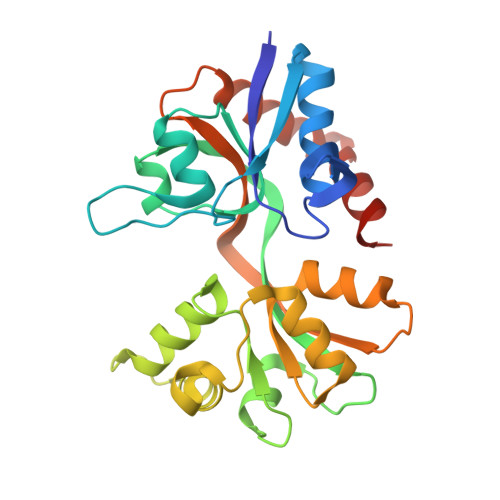
The GluD2 receptor is a fundamental component of postsynaptic sites in Purkinje neurons, and is required for normal cerebellar function. GluD2 and the closely related GluD1 are classified as members of the ionotropic glutamate receptor (iGluR) superfamily on the basis of sequence similarity, but do not bind l-glutamate. The amino acid neurotransmitter D-Ser is a GluD2 receptor ligand, and endogenous D-Ser signaling through GluD2 has recently been shown to regulate endocytosis of α-amino-3-hydroxy-5-methyl-4-isoxazolepropionic acid-type iGluRs during synaptic plasticity in the cerebellum, such as long-term depression. Here, we investigate the pharmacology of the orthosteric binding site in GluD2 by examining the activity of analogs of D-Ser and GluN1 glycine site competitive antagonists at GluD2 receptors containing the lurcher mutation (GluD2(LC)), which promotes spontaneous channel activation. We identify several compounds that modulate GluD2(LC), including a halogenated alanine analog as well as the kynurenic acid analog 7-chloro-4-oxo-1H-quinoline-2-carboxylic acid (7-chlorokynurenic acid; 7-CKA). By correlating thermodynamic and structural data for 7-CKA binding to the isolated GluD2 ligand binding domain (GluD2-LBD), we find that binding 7-CKA to GluD2-LBD differs from D-Ser by inducing an intermediate cleft closure of the clamshell-shaped LBD. The GluD2 ligands identified here can potentially serve as a starting point for development of GluD2-selective ligands useful as tools in studies of the signaling role of the GluD2 receptor in the brain.
Organizational Affiliation:
Department of Pharmacology, Emory University School of Medicine, Atlanta, Georgia (A.S.K., K.B.H., N.L.K., S.M.D., S.F.T.); Department of Drug Design and Pharmacology, University of Copenhagen, Copenhagen, Denmark (A.S.K., P.N., L.O., T.K., J.P., R.P.C., M.G., J.S.K.); and Department of Biomedical and Pharmaceutical Sciences, University of Montana, Missoula, Montana (K.B.H., F.Y.).