5CBR

Crystal structure of the GluA2 ligand-binding domain (S1S2J) in complex with the antagonist (S)-2-amino-3-(3,4-dichloro-5-(5-hydroxypyridin-3-yl)phenyl)propanoic acid at 2.0A resolution


Experimental Data Snapshot

  • Method: X-RAY DIFFRACTION
  • Resolution: 2.00 Å
  • R-Value Free: 0.238 
  • R-Value Work: 0.186 
  • R-Value Observed: 0.188 

wwPDB Validation   3D Report Full Report


Ligand Structure Quality Assessment 


This is version 1.3 of the entry. See complete history


Literature

Studies on Aryl-Substituted Phenylalanines: Synthesis, Activity, and Different Binding Modes at AMPA Receptors.

Szymanska, E.Frydenvang, K.Pickering, D.S.Krintel, C.Nielsen, B.Kooshki, A.Zachariassen, L.G.Olsen, L.Kastrup, J.S.Johansen, T.N.

(2016) J Med Chem 59: 448-461

  • DOI: https://doi.org/10.1021/acs.jmedchem.5b01666
  • Primary Citation of Related Structures:  
    5CBR, 5CBS

  • PubMed Abstract: 

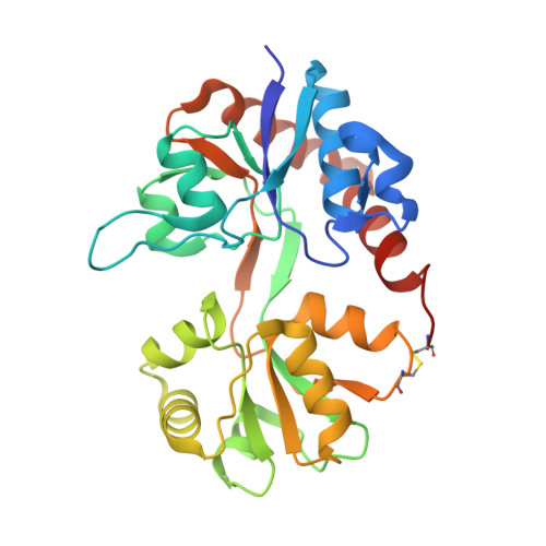
    A series of racemic aryl-substituted phenylalanines was synthesized and evaluated in vitro at recombinant rat GluA1-3, at GluK1-3, and at native AMPA receptors. The individual enantiomers of two target compounds, (RS)-2-amino-3-(3,4-dichloro-5-(5-hydroxypyridin-3-yl)phenyl)propanoic acid 37 and (RS)-2-amino-3-(3'-hydroxybiphenyl-3-yl)propanoic acid 38, were characterized. (S)-37 and (R)-38 were identified as the only biologically active isomers, both being antagonists at GluA2 receptors with Kb of 1.80 and 3.90 μM, respectively. To address this difference in enantiopharmacology, not previously seen for amino acid-based AMPA receptor antagonists, X-ray crystal structures of both eutomers in complex with the GluA2 ligand binding domain were solved. The cocrystal structures of (S)-37 and (R)-38 showed similar interactions of the amino acid parts but unexpected and different orientations and interactions of the biaromatic parts of the ligands inside the binding site, with (R)-38 having a binding mode not previously identified for amino acid-based antagonists.


  • Organizational Affiliation

    Department of Drug Design and Pharmacology, Faculty of Health and Medical Sciences, University of Copenhagen , 2100 Copenhagen, Denmark.


Macromolecules
Find similar proteins by:  (by identity cutoff)  |  3D Structure
Entity ID: 1
MoleculeChains Sequence LengthOrganismDetailsImage
Glutamate receptor 2,Glutamate receptor 2264Rattus norvegicusMutation(s): 0 
Gene Names: Gria2Glur2
UniProt
Find proteins for P19491 (Rattus norvegicus)
Explore P19491 
Go to UniProtKB:  P19491
Entity Groups  
Sequence Clusters30% Identity50% Identity70% Identity90% Identity95% Identity100% Identity
UniProt GroupP19491
Sequence Annotations
Expand
  • Reference Sequence
Small Molecules
Ligands 4 Unique
IDChains Name / Formula / InChI Key2D Diagram3D Interactions
4ZK
Query on 4ZK

Download Ideal Coordinates CCD File 
B [auth A]3,4-dichloro-5-(5-hydroxypyridin-3-yl)-L-phenylalanine
C14 H12 Cl2 N2 O3
JDADEROVQGCQQJ-LBPRGKRZSA-N
SO4
Query on SO4

Download Ideal Coordinates CCD File 
F [auth A]
G [auth A]
H [auth A]
I [auth A]
J [auth A]
F [auth A],
G [auth A],
H [auth A],
I [auth A],
J [auth A],
K [auth A]
SULFATE ION
O4 S
QAOWNCQODCNURD-UHFFFAOYSA-L
GOL
Query on GOL

Download Ideal Coordinates CCD File 
C [auth A],
D [auth A],
E [auth A]
GLYCEROL
C3 H8 O3
PEDCQBHIVMGVHV-UHFFFAOYSA-N
ACT
Query on ACT

Download Ideal Coordinates CCD File 
L [auth A]ACETATE ION
C2 H3 O2
QTBSBXVTEAMEQO-UHFFFAOYSA-M
Binding Affinity Annotations 
IDSourceBinding Affinity
4ZK BindingDB:  5CBR Ki: min: 1740, max: 6880 (nM) from 3 assay(s)
IC50: 3700 (nM) from 1 assay(s)
Binding MOAD:  5CBR Kd: 1800 (nM) from 1 assay(s)
Experimental Data & Validation

Experimental Data

  • Method: X-RAY DIFFRACTION
  • Resolution: 2.00 Å
  • R-Value Free: 0.238 
  • R-Value Work: 0.186 
  • R-Value Observed: 0.188 
  • Space Group: P 21 21 2
Unit Cell:
Length ( Å )Angle ( ˚ )
a = 60.13α = 90
b = 95.78β = 90
c = 49.42γ = 90
Software Package:
Software NamePurpose
SCALAdata scaling
PHENIXrefinement
PDB_EXTRACTdata extraction
MOSFLMdata reduction
PHASERphasing
Cootmodel building

Structure Validation

View Full Validation Report


Ligand Structure Quality Assessment 


Entry History 

Deposition Data

Revision History  (Full details and data files)

  • Version 1.0: 2015-12-30
    Type: Initial release
  • Version 1.1: 2016-01-13
    Changes: Database references
  • Version 1.2: 2016-01-27
    Changes: Database references
  • Version 1.3: 2024-01-10
    Changes: Data collection, Database references, Refinement description