5C5K

Structure of the Pfr form of a canonical phytochrome


Experimental Data Snapshot

  • Method: X-RAY DIFFRACTION
  • Resolution: 3.31 Å
  • R-Value Free: 0.232 
  • R-Value Work: 0.196 
  • R-Value Observed: 0.198 

wwPDB Validation   3D Report Full Report


Ligand Structure Quality Assessment 


This is version 1.5 of the entry. See complete history


Literature

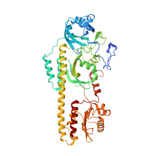
Crystal Structure of Deinococcus Phytochrome in the Photoactivated State Reveals a Cascade of Structural Rearrangements during Photoconversion.

Burgie, E.S.Zhang, J.Vierstra, R.D.

(2016) Structure 24: 448-457

  • DOI: https://doi.org/10.1016/j.str.2016.01.001
  • Primary Citation of Related Structures:  
    5C5K

  • PubMed Abstract: 

    Phytochromes are photochromic photoreceptors responsible for a myriad of red/far-red light-dependent processes in plants and microorganisms. Interconversion is initially driven by photoreversible isomerization of bilin, but how this alteration directs the photostate-dependent changes within the protein to actuate signaling is poorly understood. Here, we describe the structure of the Deinococcus phytochrome photosensory module in its near complete far-red light-absorbing Pfr state. In addition to confirming the 180° rotation of the D-pyrrole ring, the dimeric structure clearly identifies downstream rearrangements that trigger large-scale conformational differences between the dark-adapted and photoactivated states. Mutational analyses verified the importance of residues surrounding the bilin in Pfr stabilization, and protease sensitivity assays corroborated photostate alterations that propagate along the dimeric interface. Collectively, these data support a cooperative "toggle" model for phytochrome photoconversion and advance our understanding of the allosteric connection between the photosensory and output modules.


  • Organizational Affiliation

    Department of Genetics, University of Wisconsin-Madison, 425-G Henry Mall, Madison, WI 53706, USA; Department of Biology, Washington University in St Louis, Campus Box 1137, One Brookings Drive, St. Louis, MO 63130, USA.


Macromolecules
Find similar proteins by:  (by identity cutoff)  |  3D Structure
Entity ID: 1
MoleculeChains Sequence LengthOrganismDetailsImage
Bacteriophytochrome
A, B, C, D
524Deinococcus radiodurans R1 = ATCC 13939 = DSM 20539Mutation(s): 1 
Gene Names: bphPDR_A0050
EC: 2.7.13.3
UniProt
Find proteins for Q9RZA4 (Deinococcus radiodurans (strain ATCC 13939 / DSM 20539 / JCM 16871 / CCUG 27074 / LMG 4051 / NBRC 15346 / NCIMB 9279 / VKM B-1422 / R1))
Explore Q9RZA4 
Go to UniProtKB:  Q9RZA4
Entity Groups  
Sequence Clusters30% Identity50% Identity70% Identity90% Identity95% Identity100% Identity
UniProt GroupQ9RZA4
Sequence Annotations
Expand
  • Reference Sequence
Small Molecules
Ligands 4 Unique
IDChains Name / Formula / InChI Key2D Diagram3D Interactions
BLA
Query on BLA

Download Ideal Coordinates CCD File 
E [auth A],
H [auth B],
I [auth C],
K [auth D]
BILIVERDINE IX ALPHA
C33 H34 N4 O6
GWZYPXHJIZCRAJ-SRVCBVSDSA-N
BGC
Query on BGC

Download Ideal Coordinates CCD File 
J [auth C]beta-D-glucopyranose
C6 H12 O6
WQZGKKKJIJFFOK-VFUOTHLCSA-N
EDO
Query on EDO

Download Ideal Coordinates CCD File 
F [auth A]1,2-ETHANEDIOL
C2 H6 O2
LYCAIKOWRPUZTN-UHFFFAOYSA-N
ACT
Query on ACT

Download Ideal Coordinates CCD File 
G [auth A]ACETATE ION
C2 H3 O2
QTBSBXVTEAMEQO-UHFFFAOYSA-M
Experimental Data & Validation

Experimental Data

  • Method: X-RAY DIFFRACTION
  • Resolution: 3.31 Å
  • R-Value Free: 0.232 
  • R-Value Work: 0.196 
  • R-Value Observed: 0.198 
  • Space Group: P 21 21 21
Unit Cell:
Length ( Å )Angle ( ˚ )
a = 89.399α = 90
b = 192.747β = 90
c = 225.071γ = 90
Software Package:
Software NamePurpose
PHENIXrefinement
Aimlessdata scaling
PHASERphasing
PDB_EXTRACTdata extraction
XDSdata scaling
XDSdata reduction

Structure Validation

View Full Validation Report


Ligand Structure Quality Assessment 


Entry History & Funding Information

Deposition Data


Funding OrganizationLocationGrant Number
National Science Foundation (NSF, United States)United StatesMCB-1329956

Revision History  (Full details and data files)

  • Version 1.0: 2016-02-17
    Type: Initial release
  • Version 1.1: 2016-03-16
    Changes: Database references
  • Version 1.2: 2017-09-20
    Changes: Author supporting evidence, Database references, Derived calculations
  • Version 1.3: 2019-11-27
    Changes: Author supporting evidence, Data collection
  • Version 1.4: 2020-07-29
    Type: Remediation
    Reason: Carbohydrate remediation
    Changes: Data collection, Derived calculations, Structure summary
  • Version 1.5: 2023-09-27
    Changes: Data collection, Database references, Refinement description, Structure summary