5C33

Crystal Structure of Mouse Ryanodine Receptor 2 SPRY1 Domain

  • Classification: CONTRACTILE PROTEIN
  • Organism(s): Mus musculus
  • Expression System: Escherichia coli BL21(DE3)
  • Mutation(s): No 
  • Membrane Protein: Yes  mpstruc

  • Deposited: 2015-06-16 Released: 2015-08-05 
  • Deposition Author(s): Yuchi, Z., Van Petegem, F.
  • Funding Organization(s): Canadian Institutes of Health Research (CIHR), National Institutes of Health/National Institute of Arthritis and Musculoskeletal and Skin Diseases (NIH/NIAMS), National Institutes of Health/National Heart, Lung, and Blood Institute (NIH/NHLBI)

Experimental Data Snapshot

  • Method: X-RAY DIFFRACTION
  • Resolution: 1.21 Å
  • R-Value Free: 0.172 
  • R-Value Work: 0.143 
  • R-Value Observed: 0.145 

wwPDB Validation   3D Report Full Report


This is version 1.3 of the entry. See complete history


Literature

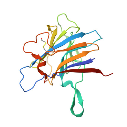
Crystal structures of ryanodine receptor SPRY1 and tandem-repeat domains reveal a critical FKBP12 binding determinant.

Yuchi, Z.Yuen, S.M.Lau, K.Underhill, A.Q.Cornea, R.L.Fessenden, J.D.Van Petegem, F.

(2015) Nat Commun 6: 7947-7947

  • DOI: https://doi.org/10.1038/ncomms8947
  • Primary Citation of Related Structures:  
    5C30, 5C33

  • PubMed Abstract: 

    Ryanodine receptors (RyRs) form calcium release channels located in the membranes of the sarcoplasmic and endoplasmic reticulum. RyRs play a major role in excitation-contraction coupling and other Ca(2+)-dependent signalling events, and consist of several globular domains that together form a large assembly. Here we describe the crystal structures of the SPRY1 and tandem-repeat domains at 1.2-1.5 Å resolution, which reveal several structural elements not detected in recent cryo-EM reconstructions of RyRs. The cryo-EM studies disagree on the position of SPRY domains, which had been proposed based on homology modelling. Computational docking of the crystal structures, combined with FRET studies, show that the SPRY1 domain is located next to FK506-binding protein (FKBP). Molecular dynamics flexible fitting and mutagenesis experiments suggest a hydrophobic cluster within SPRY1 that is crucial for FKBP binding. A RyR1 disease mutation, N760D, appears to directly impact FKBP binding through interfering with SPRY1 folding.


  • Organizational Affiliation

    Department of Biochemistry and Molecular Biology, The Life Sciences Centre, University of British Columbia, 2350 Health Sciences Mall, Vancouver, Canada V6T 1Z3.


Macromolecules
Find similar proteins by:  (by identity cutoff)  |  3D Structure
Entity ID: 1
MoleculeChains Sequence LengthOrganismDetailsImage
Ryanodine receptor 2
A, B
198Mus musculusMutation(s): 0 
Gene Names: Ryr2
Membrane Entity: Yes 
UniProt & NIH Common Fund Data Resources
Find proteins for E9Q401 (Mus musculus)
Explore E9Q401 
Go to UniProtKB:  E9Q401
IMPC:  MGI:99685
Entity Groups  
Sequence Clusters30% Identity50% Identity70% Identity90% Identity95% Identity100% Identity
UniProt GroupE9Q401
Sequence Annotations
Expand
  • Reference Sequence
Small Molecules
Modified Residues  1 Unique
IDChains TypeFormula2D DiagramParent
CME
Query on CME
A, B
L-PEPTIDE LINKINGC5 H11 N O3 S2CYS
Experimental Data & Validation

Experimental Data

  • Method: X-RAY DIFFRACTION
  • Resolution: 1.21 Å
  • R-Value Free: 0.172 
  • R-Value Work: 0.143 
  • R-Value Observed: 0.145 
  • Space Group: P 21 21 21
Unit Cell:
Length ( Å )Angle ( ˚ )
a = 53.246α = 90
b = 64.085β = 90
c = 109.769γ = 90
Software Package:
Software NamePurpose
PHENIXrefinement
XDSdata reduction
XDSdata scaling
PHENIXphasing

Structure Validation

View Full Validation Report


Entry History & Funding Information

Deposition Data


Funding OrganizationLocationGrant Number
Canadian Institutes of Health Research (CIHR)CanadaMOP-119608
National Institutes of Health/National Institute of Arthritis and Musculoskeletal and Skin Diseases (NIH/NIAMS)United StatesR01AR059124
National Institutes of Health/National Heart, Lung, and Blood Institute (NIH/NHLBI)United StatesR01HL092097

Revision History  (Full details and data files)

  • Version 1.0: 2015-08-05
    Type: Initial release
  • Version 1.1: 2015-08-19
    Changes: Database references
  • Version 1.2: 2017-09-06
    Changes: Author supporting evidence, Derived calculations
  • Version 1.3: 2019-12-04
    Changes: Author supporting evidence