5C1C

Crystal Structure of the Pectin Methylesterase from Aspergillus niger in Deglycosylated Form


Experimental Data Snapshot

  • Method: X-RAY DIFFRACTION
  • Resolution: 1.80 Å
  • R-Value Free: 0.203 
  • R-Value Work: 0.170 
  • R-Value Observed: 0.172 

wwPDB Validation   3D Report Full Report


This is version 1.4 of the entry. See complete history


Literature

Structure and Properties of a Non-processive, Salt-requiring, and Acidophilic Pectin Methylesterase from Aspergillus niger Provide Insights into the Key Determinants of Processivity Control.

Kent, L.M.Loo, T.S.Melton, L.D.Mercadante, D.Williams, M.A.Jameson, G.B.

(2016) J Biol Chem 291: 1289-1306

  • DOI: https://doi.org/10.1074/jbc.M115.673152
  • Primary Citation of Related Structures:  
    5C1C

  • PubMed Abstract: 

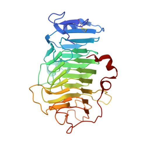
    Many pectin methylesterases (PMEs) are expressed in plants to modify plant cell-wall pectins for various physiological roles. These pectins are also attacked by PMEs from phytopathogens and phytophagous insects. The de-methylesterification by PMEs of the O6-methyl ester groups of the homogalacturonan component of pectin, exposing galacturonic acids, can occur processively or non-processively, respectively, describing sequential versus single de-methylesterification events occurring before enzyme-substrate dissociation. The high resolution x-ray structures of a PME from Aspergillus niger in deglycosylated and Asn-linked N-acetylglucosamine-stub forms reveal a 10⅔-turn parallel β-helix (similar to but with less extensive loops than bacterial, plant, and insect PMEs). Capillary electrophoresis shows that this PME is non-processive, halophilic, and acidophilic. Molecular dynamics simulations and electrostatic potential calculations reveal very different behavior and properties compared with processive PMEs. Specifically, uncorrelated rotations are observed about the glycosidic bonds of a partially de-methyl-esterified decasaccharide model substrate, in sharp contrast to the correlated rotations of processive PMEs, and the substrate-binding groove is negatively not positively charged.


  • Organizational Affiliation

    From Riddet Institute and Institute of Fundamental Sciences, Massey University, Palmerston North 4442, New Zealand.


Macromolecules
Find similar proteins by:  (by identity cutoff)  |  3D Structure
Entity ID: 1
MoleculeChains Sequence LengthOrganismDetailsImage
Pectinesterase299Aspergillus niger ATCC 1015Mutation(s): 1 
Gene Names: ASPNIDRAFT_214857
EC: 3.1.1.11
UniProt
Find proteins for G3YAL0 (Aspergillus niger (strain ATCC 1015 / CBS 113.46 / FGSC A1144 / LSHB Ac4 / NCTC 3858a / NRRL 328 / USDA 3528.7))
Explore G3YAL0 
Go to UniProtKB:  G3YAL0
Entity Groups  
Sequence Clusters30% Identity50% Identity70% Identity90% Identity95% Identity100% Identity
UniProt GroupG3YAL0
Sequence Annotations
Expand
  • Reference Sequence
Small Molecules
Ligands 4 Unique
IDChains Name / Formula / InChI Key2D Diagram3D Interactions
SO4
Query on SO4

Download Ideal Coordinates CCD File 
B [auth A],
C [auth A],
D [auth A],
E [auth A],
F [auth A]
SULFATE ION
O4 S
QAOWNCQODCNURD-UHFFFAOYSA-L
GOL
Query on GOL

Download Ideal Coordinates CCD File 
H [auth A],
I [auth A],
J [auth A]
GLYCEROL
C3 H8 O3
PEDCQBHIVMGVHV-UHFFFAOYSA-N
ACT
Query on ACT

Download Ideal Coordinates CCD File 
K [auth A]ACETATE ION
C2 H3 O2
QTBSBXVTEAMEQO-UHFFFAOYSA-M
CL
Query on CL

Download Ideal Coordinates CCD File 
G [auth A]CHLORIDE ION
Cl
VEXZGXHMUGYJMC-UHFFFAOYSA-M
Experimental Data & Validation

Experimental Data

  • Method: X-RAY DIFFRACTION
  • Resolution: 1.80 Å
  • R-Value Free: 0.203 
  • R-Value Work: 0.170 
  • R-Value Observed: 0.172 
  • Space Group: C 2 2 21
Unit Cell:
Length ( Å )Angle ( ˚ )
a = 75.249α = 90
b = 113.843β = 90
c = 88.741γ = 90
Software Package:
Software NamePurpose
d*TREKdata scaling
REFMACrefinement
PDB_EXTRACTdata extraction
CrystalCleardata reduction
MrBUMPphasing

Structure Validation

View Full Validation Report


Entry History 

Deposition Data

Revision History  (Full details and data files)

  • Version 1.0: 2015-07-01
    Type: Initial release
  • Version 1.1: 2015-12-02
    Changes: Database references
  • Version 1.2: 2016-02-03
    Changes: Database references
  • Version 1.3: 2017-11-01
    Changes: Author supporting evidence, Database references, Derived calculations, Refinement description
  • Version 1.4: 2023-09-27
    Changes: Data collection, Database references, Refinement description