5BTR

Crystal structure of SIRT1 in complex with resveratrol and an AMC-containing peptide


Experimental Data Snapshot

  • Method: X-RAY DIFFRACTION
  • Resolution: 3.20 Å
  • R-Value Free: 0.252 
  • R-Value Work: 0.207 
  • R-Value Observed: 0.209 

wwPDB Validation   3D Report Full Report


Ligand Structure Quality Assessment 


This is version 1.3 of the entry. See complete history


Literature

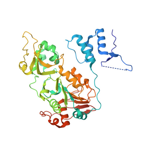
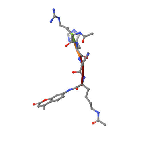
Structural basis for allosteric, substrate-dependent stimulation of SIRT1 activity by resveratrol

Cao, D.Wang, M.Qiu, X.Liu, D.Jiang, H.Yang, N.Xu, R.M.

(2015) Genes Dev 29: 1316-1325

  • DOI: https://doi.org/10.1101/gad.265462.115
  • Primary Citation of Related Structures:  
    5BTR

  • PubMed Abstract: 

    Sirtuins with an extended N-terminal domain (NTD), represented by yeast Sir2 and human SIRT1, harbor intrinsic mechanisms for regulation of their NAD-dependent deacetylase activities. Elucidation of the regulatory mechanisms is crucial for understanding the biological functions of sirtuins and development of potential therapeutics. In particular, SIRT1 has emerged as an attractive therapeutic target, and the search for SIRT1-activating compounds (STACs) has been actively pursued. However, the effectiveness of a class of reported STACs (represented by resveratrol) as direct SIRT1 activators is under debate due to the complication involving the use of fluorogenic substrates in in vitro assays. Future efforts of SIRT1-based therapeutics necessitate the dissection of the molecular mechanism of SIRT1 stimulation. We solved the structure of SIRT1 in complex with resveratrol and a 7-amino-4-methylcoumarin (AMC)-containing peptide. The structure reveals the presence of three resveratrol molecules, two of which mediate the interaction between the AMC peptide and the NTD of SIRT1. The two NTD-bound resveratrol molecules are principally responsible for promoting tighter binding between SIRT1 and the peptide and the stimulation of SIRT1 activity. The structural information provides valuable insights into regulation of SIRT1 activity and should benefit the development of authentic SIRT1 activators.


  • Organizational Affiliation

    National Laboratory of Biomacromolecules, Institute of Biophysics, Chinese Academy of Sciences, Beijing 100101, China; University of Chinese Academy of Sciences, Beijing 100049, China;


Macromolecules
Find similar proteins by:  (by identity cutoff)  |  3D Structure
Entity ID: 1
MoleculeChains Sequence LengthOrganismDetailsImage
NAD-dependent protein deacetylase sirtuin-1
A, B, C
397Homo sapiensMutation(s): 4 
Gene Names: SIRT1SIR2L1
EC: 3.5.1
UniProt & NIH Common Fund Data Resources
Find proteins for Q96EB6 (Homo sapiens)
Explore Q96EB6 
Go to UniProtKB:  Q96EB6
PHAROS:  Q96EB6
GTEx:  ENSG00000096717 
Entity Groups  
Sequence Clusters30% Identity50% Identity70% Identity90% Identity95% Identity100% Identity
UniProt GroupQ96EB6
Sequence Annotations
Expand
  • Reference Sequence

Find similar proteins by:  Sequence   |   3D Structure  

Entity ID: 2
MoleculeChains Sequence LengthOrganismDetailsImage
AMC-containing peptide
D, E, F
5synthetic constructMutation(s): 0 
Sequence Annotations
Expand
  • Reference Sequence
Small Molecules
Ligands 2 Unique
IDChains Name / Formula / InChI Key2D Diagram3D Interactions
STL
Query on STL

Download Ideal Coordinates CCD File 
H [auth A]
J [auth B]
K [auth B]
M [auth C]
N [auth C]
H [auth A],
J [auth B],
K [auth B],
M [auth C],
N [auth C],
O [auth D],
P [auth D],
Q [auth E],
R [auth F]
RESVERATROL
C14 H12 O3
LUKBXSAWLPMMSZ-OWOJBTEDSA-N
ZN
Query on ZN

Download Ideal Coordinates CCD File 
G [auth A],
I [auth B],
L [auth C]
ZINC ION
Zn
PTFCDOFLOPIGGS-UHFFFAOYSA-N
Modified Residues  1 Unique
IDChains TypeFormula2D DiagramParent
FDL
Query on FDL
D, E, F
L-PEPTIDE LINKINGC18 H23 N3 O4LYS
Binding Affinity Annotations 
IDSourceBinding Affinity
STL BindingDB:  5BTR EC50: min: 2.36e+4, max: 4.62e+4 (nM) from 2 assay(s)
Experimental Data & Validation

Experimental Data

  • Method: X-RAY DIFFRACTION
  • Resolution: 3.20 Å
  • R-Value Free: 0.252 
  • R-Value Work: 0.207 
  • R-Value Observed: 0.209 
  • Space Group: P 41
Unit Cell:
Length ( Å )Angle ( ˚ )
a = 133.89α = 90
b = 133.89β = 90
c = 106.707γ = 90
Software Package:
Software NamePurpose
PHENIXrefinement
HKL-2000data collection
HKL-2000data scaling
PHASERphasing
PDB_EXTRACTdata extraction

Structure Validation

View Full Validation Report


Ligand Structure Quality Assessment 


Entry History & Funding Information

Deposition Data


Funding OrganizationLocationGrant Number
Ministry of Science and Technology (China)China2012CB910702
National Natural Science Foundation of ChinaChina31430018
National Natural Science Foundation of ChinaChina31210103914
Strategic Priority Research Program of Chinese Academy of SciencesChinaXDB08010100
Key Research Program of Chinese Academy of SciencesChinaKJZD-EW-L05
National Key New Drug Creation and Manufacturing Program of ChinaChina2014ZX09507002

Revision History  (Full details and data files)

  • Version 1.0: 2015-07-08
    Type: Initial release
  • Version 1.1: 2016-12-28
    Changes: Other
  • Version 1.2: 2017-10-18
    Changes: Author supporting evidence
  • Version 1.3: 2023-11-08
    Changes: Data collection, Database references, Derived calculations, Refinement description