5BSZ

X-ray structure of the sugar N-methyltransferase KedS8 from Streptoalloteichus sp ATCC 53650


Experimental Data Snapshot

  • Method: X-RAY DIFFRACTION
  • Resolution: 2.00 Å
  • R-Value Free: 0.226 
  • R-Value Work: 0.168 
  • R-Value Observed: 0.171 

wwPDB Validation   3D Report Full Report


Ligand Structure Quality Assessment 


This is version 1.2 of the entry. See complete history


Literature

Molecular architecture of KedS8, a sugar N-methyltransferase from Streptoalloteichus sp. ATCC 53650.

Delvaux, N.A.Thoden, J.B.Holden, H.M.

(2015) Protein Sci 24: 1593-1599

  • DOI: https://doi.org/10.1002/pro.2742
  • Primary Citation of Related Structures:  
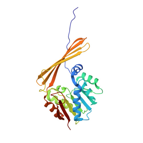
    5BSZ

  • PubMed Abstract: 

    Kedarcidin, produced by Streptoalloteichus sp. ATCC 53650, is a fascinating chromoprotein of 114 amino acid residues that displays both antibiotic and anticancer activity. The chromophore responsible for its chemotherapeutic activity is an ansa-bridged enediyne with two attached sugars, l-mycarose, and l-kedarosamine. The biosynthesis of l-kedarosamine, a highly unusual trideoxysugar, is beginning to be revealed through bioinformatics approaches. One of the enzymes putatively involved in the production of this carbohydrate is referred to as KedS8. It has been proposed that KedS8 is an N-methyltransferase that utilizes S-adenosylmethionine as the methyl donor and a dTDP-linked C-4' amino sugar as the substrate. Here we describe the three-dimensional architecture of KedS8 in complex with S-adenosylhomocysteine. The structure was solved to 2.0 Å resolution and refined to an overall R-factor of 17.1%. Unlike that observed for other sugar N-methyltransferases, KedS8 adopts a novel tetrameric quaternary structure due to the swapping of β-strands at the N-termini of its subunits. The structure presented here represents the first example of an N-methyltransferase that functions on C-4' rather than C-3' amino sugars.


  • Organizational Affiliation

    Department of Biochemistry, University of Wisconsin, Madison, Wisconsin, 53706.


Macromolecules
Find similar proteins by:  (by identity cutoff)  |  3D Structure
Entity ID: 1
MoleculeChains Sequence LengthOrganismDetailsImage
N-methyltransferase250Streptoalloteichus sp. ATCC 53650Mutation(s): 0 
UniProt
Find proteins for K4P132 (Streptoalloteichus sp. ATCC 53650)
Explore K4P132 
Go to UniProtKB:  K4P132
Entity Groups  
Sequence Clusters30% Identity50% Identity70% Identity90% Identity95% Identity100% Identity
UniProt GroupK4P132
Sequence Annotations
Expand
  • Reference Sequence
Experimental Data & Validation

Experimental Data

  • Method: X-RAY DIFFRACTION
  • Resolution: 2.00 Å
  • R-Value Free: 0.226 
  • R-Value Work: 0.168 
  • R-Value Observed: 0.171 
  • Space Group: I 2 2 2
Unit Cell:
Length ( Å )Angle ( ˚ )
a = 63.254α = 90
b = 81.004β = 90
c = 126.916γ = 90
Software Package:
Software NamePurpose
REFMACrefinement
SAINTdata reduction
SADABSdata scaling
PHASERphasing

Structure Validation

View Full Validation Report


Ligand Structure Quality Assessment 


Entry History 

Deposition Data

Revision History  (Full details and data files)

  • Version 1.0: 2015-07-29
    Type: Initial release
  • Version 1.1: 2015-10-14
    Changes: Database references
  • Version 1.2: 2023-09-27
    Changes: Data collection, Database references, Derived calculations, Refinement description