5BS7

Structure of histone H3/H4 in complex with Spt2

  • Classification: TRANSCRIPTION REGULATOR
  • Organism(s): Xenopus laevis, Homo sapiens
  • Expression System: Escherichia coli
  • Mutation(s): No 

  • Deposited: 2015-06-01 Released: 2015-07-08 
  • Deposition Author(s): Chen, S., Patel, D.J.
  • Funding Organization(s): Leukemia & Lymphoma Society, STARR Foundation grant, National Institutes of Health/National Institute of General Medical Sciences (NIH/NIGMS), National Institutes of Health/National Center for Research Resources (NIH/NCRR)

Experimental Data Snapshot

  • Method: X-RAY DIFFRACTION
  • Resolution: 3.30 Å
  • R-Value Free: 0.290 
  • R-Value Work: 0.230 
  • R-Value Observed: 0.233 

wwPDB Validation   3D Report Full Report


This is version 1.5 of the entry. See complete history


Literature

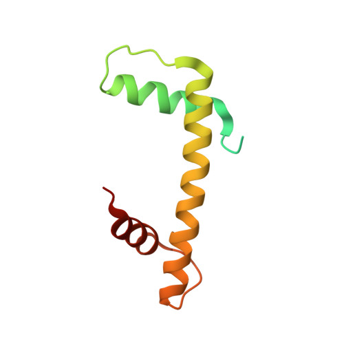
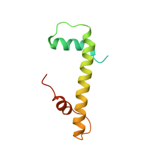
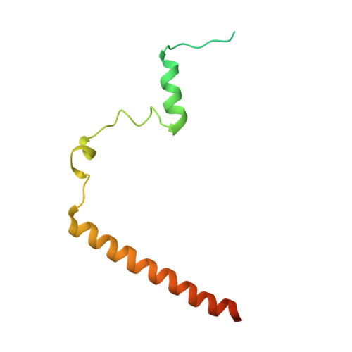
Structure-function studies of histone H3/H4 tetramer maintenance during transcription by chaperone Spt2.

Chen, S.Rufiange, A.Huang, H.Rajashankar, K.R.Nourani, A.Patel, D.J.

(2015) Genes Dev 29: 1326-1340

  • DOI: https://doi.org/10.1101/gad.261115.115
  • Primary Citation of Related Structures:  
    5BS7, 5BSA

  • PubMed Abstract: 

    Cells use specific mechanisms such as histone chaperones to abrogate the inherent barrier that the nucleosome poses to transcribing polymerases. The current model postulates that nucleosomes can be transiently disrupted to accommodate passage of RNA polymerases and that histones H3 and H4 possess their own chaperones dedicated to the recovery of nucleosomes. Here, we determined the crystal structure of the conserved C terminus of human Suppressors of Ty insertions 2 (hSpt2C) chaperone bound to an H3/H4 tetramer. The structural studies demonstrate that hSpt2C is bound to the periphery of the H3/H4 tetramer, mimicking the trajectory of nucleosomal-bound DNA. These structural studies have been complemented with in vitro binding and in vivo functional studies on mutants that disrupt key intermolecular contacts involving two acidic patches and hydrophobic residues on Spt2C. We show that contacts between both human and yeast Spt2C with the H3/H4 tetramer are required for the suppression of H3/H4 exchange as measured by H3K56ac and new H3 deposition. These interactions are also crucial for the inhibition of spurious transcription from within coding regions. Together, our data indicate that Spt2 interacts with the periphery of the H3/H4 tetramer and promotes its recycling in the wake of RNA polymerase.


  • Organizational Affiliation

    Structural Biology Program, Memorial Sloan-Kettering Cancer Center, New York, New York 10065, USA;


Macromolecules
Find similar proteins by:  (by identity cutoff)  |  3D Structure
Entity ID: 1
MoleculeChains Sequence LengthOrganismDetailsImage
Histone H3.2
A, B
111Xenopus laevisMutation(s): 0 
UniProt
Find proteins for P84233 (Xenopus laevis)
Explore P84233 
Go to UniProtKB:  P84233
Entity Groups  
Sequence Clusters30% Identity50% Identity70% Identity90% Identity95% Identity100% Identity
UniProt GroupP84233
Sequence Annotations
Expand
  • Reference Sequence
Find similar proteins by:  (by identity cutoff)  |  3D Structure
Entity ID: 2
MoleculeChains Sequence LengthOrganismDetailsImage
Histone H4
C, D
102Xenopus laevisMutation(s): 0 
UniProt
Find proteins for P62799 (Xenopus laevis)
Explore P62799 
Go to UniProtKB:  P62799
Entity Groups  
Sequence Clusters30% Identity50% Identity70% Identity90% Identity95% Identity100% Identity
UniProt GroupP62799
Sequence Annotations
Expand
  • Reference Sequence
Find similar proteins by:  (by identity cutoff)  |  3D Structure
Entity ID: 3
MoleculeChains Sequence LengthOrganismDetailsImage
Protein SPT2 homolog
E, F
115Homo sapiensMutation(s): 0 
Gene Names: SPTY2D1
UniProt & NIH Common Fund Data Resources
Find proteins for Q68D10 (Homo sapiens)
Explore Q68D10 
Go to UniProtKB:  Q68D10
PHAROS:  Q68D10
GTEx:  ENSG00000179119 
Entity Groups  
Sequence Clusters30% Identity50% Identity70% Identity90% Identity95% Identity100% Identity
UniProt GroupQ68D10
Sequence Annotations
Expand
  • Reference Sequence
Small Molecules
Ligands 1 Unique
IDChains Name / Formula / InChI Key2D Diagram3D Interactions
SO4
Query on SO4

Download Ideal Coordinates CCD File 
G [auth C]SULFATE ION
O4 S
QAOWNCQODCNURD-UHFFFAOYSA-L
Experimental Data & Validation

Experimental Data

  • Method: X-RAY DIFFRACTION
  • Resolution: 3.30 Å
  • R-Value Free: 0.290 
  • R-Value Work: 0.230 
  • R-Value Observed: 0.233 
  • Space Group: P 43 2 2
Unit Cell:
Length ( Å )Angle ( ˚ )
a = 121.205α = 90
b = 121.205β = 90
c = 118.503γ = 90
Software Package:
Software NamePurpose
REFMACrefinement
PHASERphasing
HKL-2000data scaling
Cootmodel building
HKL-2000data reduction

Structure Validation

View Full Validation Report


Entry History & Funding Information

Deposition Data


Funding OrganizationLocationGrant Number
Leukemia & Lymphoma SocietyUnited StatesLLS-SCOR 7132-08
STARR Foundation grantUnited StatesI5-A554
National Institutes of Health/National Institute of General Medical Sciences (NIH/NIGMS)United StatesP41 GM103403
National Institutes of Health/National Center for Research Resources (NIH/NCRR)United StatesS10 RR029205

Revision History  (Full details and data files)

  • Version 1.0: 2015-07-08
    Type: Initial release
  • Version 1.1: 2017-09-20
    Changes: Author supporting evidence, Database references, Derived calculations, Refinement description, Source and taxonomy
  • Version 1.2: 2017-09-27
    Changes: Author supporting evidence
  • Version 1.3: 2019-12-25
    Changes: Author supporting evidence
  • Version 1.4: 2022-03-23
    Changes: Author supporting evidence, Database references
  • Version 1.5: 2023-09-27
    Changes: Data collection, Refinement description