5BQ1

Capturing Carbon Dioxide in beta Carbonic Anhydrase


Experimental Data Snapshot

  • Method: X-RAY DIFFRACTION
  • Resolution: 1.60 Å
  • R-Value Free: 0.199 
  • R-Value Work: 0.173 
  • R-Value Observed: 0.175 

wwPDB Validation   3D Report Full Report


This is version 1.5 of the entry. See complete history


Literature

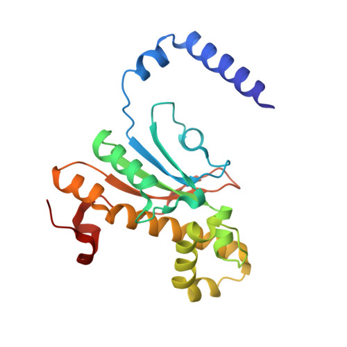
Carbon Dioxide "Trapped" in a beta-Carbonic Anhydrase.

Aggarwal, M.Chua, T.K.Pinard, M.A.Szebenyi, D.M.McKenna, R.

(2015) Biochemistry 54: 6631-6638

  • DOI: https://doi.org/10.1021/acs.biochem.5b00987
  • Primary Citation of Related Structures:  
    5BQ1

  • PubMed Abstract: 

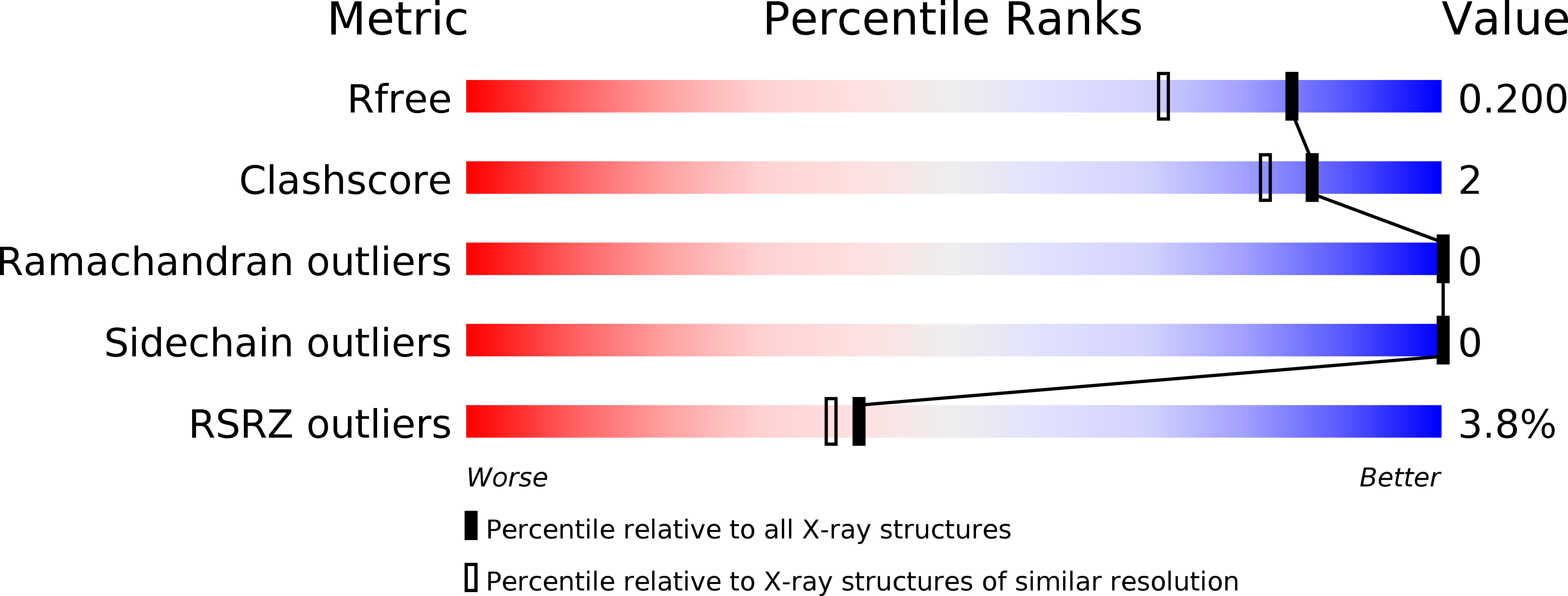
    Carbonic anhydrases (CAs) are enzymes that catalyze the hydration/dehydration of CO2/HCO3(-) with rates approaching diffusion-controlled limits (kcat/KM ∼ 10(8) M(-1) s(-1)). This family of enzymes has evolved disparate protein folds that all perform the same reaction at near catalytic perfection. Presented here is a structural study of a β-CA (psCA3) expressed in Pseudomonas aeruginosa, in complex with CO2, using pressurized cryo-cooled crystallography. The structure has been refined to 1.6 Å resolution with R(cryst) and R(free) values of 17.3 and 19.9%, respectively, and is compared with the α-CA, human CA isoform II (hCA II), the only other CA to have CO2 captured in its active site. Despite the lack of structural similarity between psCA3 and hCA II, the CO2 binding orientation relative to the zinc-bound solvent is identical. In addition, a second CO2 binding site was located at the dimer interface of psCA3. Interestingly, all β-CAs function as dimers or higher-order oligomeric states, and the CO2 bound at the interface may contribute to the allosteric nature of this family of enzymes or may be a convenient alternative binding site as this pocket has been previously shown to be a promiscuous site for a variety of ligands, including bicarbonate, sulfate, and phosphate ions.


  • Organizational Affiliation

    Division of Biology and Soft Matter, Oak Ridge National Laboratory , Oak Ridge, Tennessee 37831, United States.


Macromolecules
Find similar proteins by:  (by identity cutoff)  |  3D Structure
Entity ID: 1
MoleculeChains Sequence LengthOrganismDetailsImage
Carbonic anhydrase209Pseudomonas aeruginosaMutation(s): 0 
Gene Names: PAMH19_3356
EC: 4.2.1.1
UniProt
Find proteins for Q9HVB9 (Pseudomonas aeruginosa (strain ATCC 15692 / DSM 22644 / CIP 104116 / JCM 14847 / LMG 12228 / 1C / PRS 101 / PAO1))
Explore Q9HVB9 
Go to UniProtKB:  Q9HVB9
Entity Groups  
Sequence Clusters30% Identity50% Identity70% Identity90% Identity95% Identity100% Identity
UniProt GroupQ9HVB9
Sequence Annotations
Expand
  • Reference Sequence
Experimental Data & Validation

Experimental Data

  • Method: X-RAY DIFFRACTION
  • Resolution: 1.60 Å
  • R-Value Free: 0.199 
  • R-Value Work: 0.173 
  • R-Value Observed: 0.175 
  • Space Group: I 2 2 2
Unit Cell:
Length ( Å )Angle ( ˚ )
a = 71.874α = 90
b = 78.114β = 90
c = 87.707γ = 90
Software Package:
Software NamePurpose
HKL-2000data collection
HKL-2000data reduction
HKL-2000data scaling
PHASERphasing
PDB_EXTRACTdata extraction
PHASERphasing
PHENIXrefinement

Structure Validation

View Full Validation Report


Entry History & Funding Information

Deposition Data


Funding OrganizationLocationGrant Number
National Institutes of Health/National Institute of General Medical Sciences (NIH/NIGMS)United StatesGM25154

Revision History  (Full details and data files)

  • Version 1.0: 2015-10-28
    Type: Initial release
  • Version 1.1: 2015-11-04
    Changes: Database references
  • Version 1.2: 2015-11-11
    Changes: Database references
  • Version 1.3: 2017-09-06
    Changes: Author supporting evidence, Database references, Derived calculations
  • Version 1.4: 2019-12-25
    Changes: Author supporting evidence
  • Version 1.5: 2024-03-06
    Changes: Data collection, Database references