5B6C

Structural Details of Ufd1 binding to p97


Experimental Data Snapshot

  • Method: X-RAY DIFFRACTION
  • Resolution: 1.55 Å
  • R-Value Free: 0.200 
  • R-Value Work: 0.158 
  • R-Value Observed: 0.160 

wwPDB Validation   3D Report Full Report


This is version 1.0 of the entry. See complete history


Literature

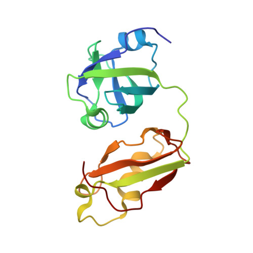
Structural Details of Ufd1 Binding to p97 and Their Functional Implications in ER-Associated Degradation

Le, L.T.M.Kang, W.Kim, J.Y.Le, O.T.T.Lee, S.Y.Yang, J.K.

(2016) PLoS One 11: e0163394-e0163394

  • DOI: https://doi.org/10.1371/journal.pone.0163394
  • Primary Citation of Related Structures:  
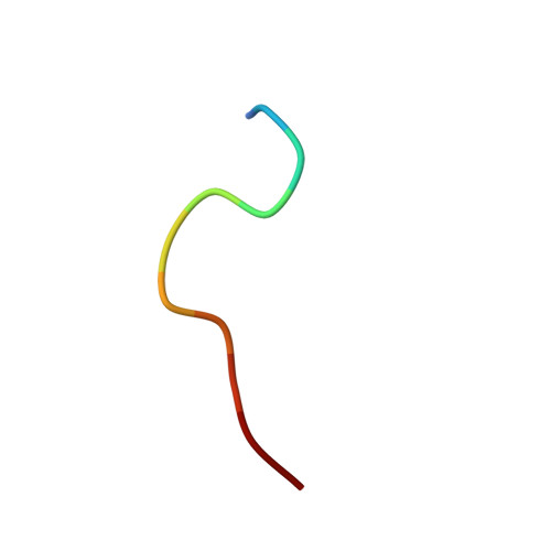
    5B6C

  • PubMed Abstract: 

    The hexameric ATPase p97 has been implicated in diverse cellular processes through interactions with many different adaptor proteins at its N-terminal domain. Among these, the Ufd1-Npl4 heterodimer is a major adaptor, and the p97-Ufd1-Npl4 complex plays an essential role in endoplasmic reticulum-associated degradation (ERAD), acting as a segregase that translocates the ubiquitinated client protein from the ER membrane into the cytosol for proteasomal degradation. We determined the crystal structure of the complex of the N-terminal domain of p97 and the SHP box of Ufd1 at a resolution of 1.55 Å. The 11-residue-long SHP box of Ufd1 binds at the far-most side of the Nc lobe of the p97 N domain primarily through hydrophobic interactions, such that F225, F228, N233 and L235 of the SHP box contact hydrophobic residues on the surface of the p97 Nc lobe. Mutating these key interface residues abolished the interactions in two different binding experiments, isothermal titration calorimetry and co-immunoprecipitation. Furthermore, cycloheximide chase assays showed that these same mutations caused accumulation of tyrosinase-C89R, a well-known ERAD substrate, thus implying decreased rate of protein degradation due to their defects in ERAD function. Together, these results provide structural and biochemical insights into the interaction between p97 N domain and Ufd1 SHP box.


  • Organizational Affiliation

    Department of Chemistry, College of Natural Sciences, Soongsil University, Seoul 156-743, Korea.


Macromolecules
Find similar proteins by:  (by identity cutoff)  |  3D Structure
Entity ID: 1
MoleculeChains Sequence LengthOrganismDetailsImage
Transitional endoplasmic reticulum ATPase174Homo sapiensMutation(s): 0 
Gene Names: VCP
EC: 3.6.4.6
UniProt & NIH Common Fund Data Resources
Find proteins for P55072 (Homo sapiens)
Explore P55072 
Go to UniProtKB:  P55072
PHAROS:  P55072
GTEx:  ENSG00000165280 
Entity Groups  
Sequence Clusters30% Identity50% Identity70% Identity90% Identity95% Identity100% Identity
UniProt GroupP55072
Sequence Annotations
Expand
  • Reference Sequence

Find similar proteins by:  Sequence   |   3D Structure  

Entity ID: 2
MoleculeChains Sequence LengthOrganismDetailsImage
Peptide from Ubiquitin fusion degradation protein 1 homolog11Homo sapiensMutation(s): 0 
Gene Names: UFD1L
UniProt & NIH Common Fund Data Resources
Find proteins for Q92890 (Homo sapiens)
Explore Q92890 
Go to UniProtKB:  Q92890
PHAROS:  Q92890
GTEx:  ENSG00000070010 
Entity Groups  
Sequence Clusters30% Identity50% Identity70% Identity90% Identity95% Identity100% Identity
UniProt GroupQ92890
Sequence Annotations
Expand
  • Reference Sequence
Experimental Data & Validation

Experimental Data

  • Method: X-RAY DIFFRACTION
  • Resolution: 1.55 Å
  • R-Value Free: 0.200 
  • R-Value Work: 0.158 
  • R-Value Observed: 0.160 
  • Space Group: H 3
Unit Cell:
Length ( Å )Angle ( ˚ )
a = 107.27α = 90
b = 107.27β = 90
c = 44.04γ = 120
Software Package:
Software NamePurpose
REFMACrefinement
MOSFLMdata reduction
SCALAdata scaling
PHASERphasing

Structure Validation

View Full Validation Report


Entry History & Funding Information

Deposition Data


Funding OrganizationLocationGrant Number
National Research Foundation of KoreaKorea, Republic OfNRF-2014R1A2A2A01006834

Revision History  (Full details and data files)

  • Version 1.0: 2017-01-04
    Type: Initial release