5B68

Crystal structure of apo amylomaltase from Corynebacterium glutamicum


Experimental Data Snapshot

  • Method: X-RAY DIFFRACTION
  • Resolution: 1.70 Å
  • R-Value Free: 0.222 
  • R-Value Work: 0.181 
  • R-Value Observed: 0.199 

wwPDB Validation   3D Report Full Report


This is version 1.1 of the entry. See complete history


Literature

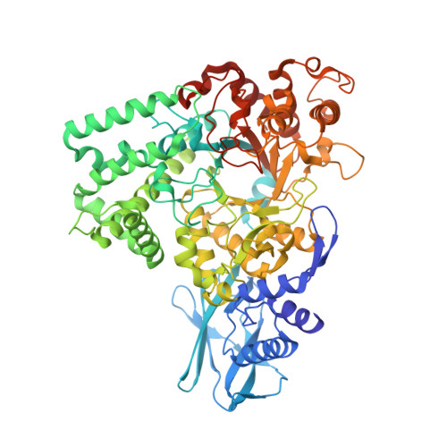
Crystal Structure of Amylomaltase from Corynebacterium glutamicum.

Joo, S.Kim, S.Seo, H.Kim, K.J.

(2016) J Agric Food Chem 64: 5662-5670

  • DOI: https://doi.org/10.1021/acs.jafc.6b02296
  • Primary Citation of Related Structures:  
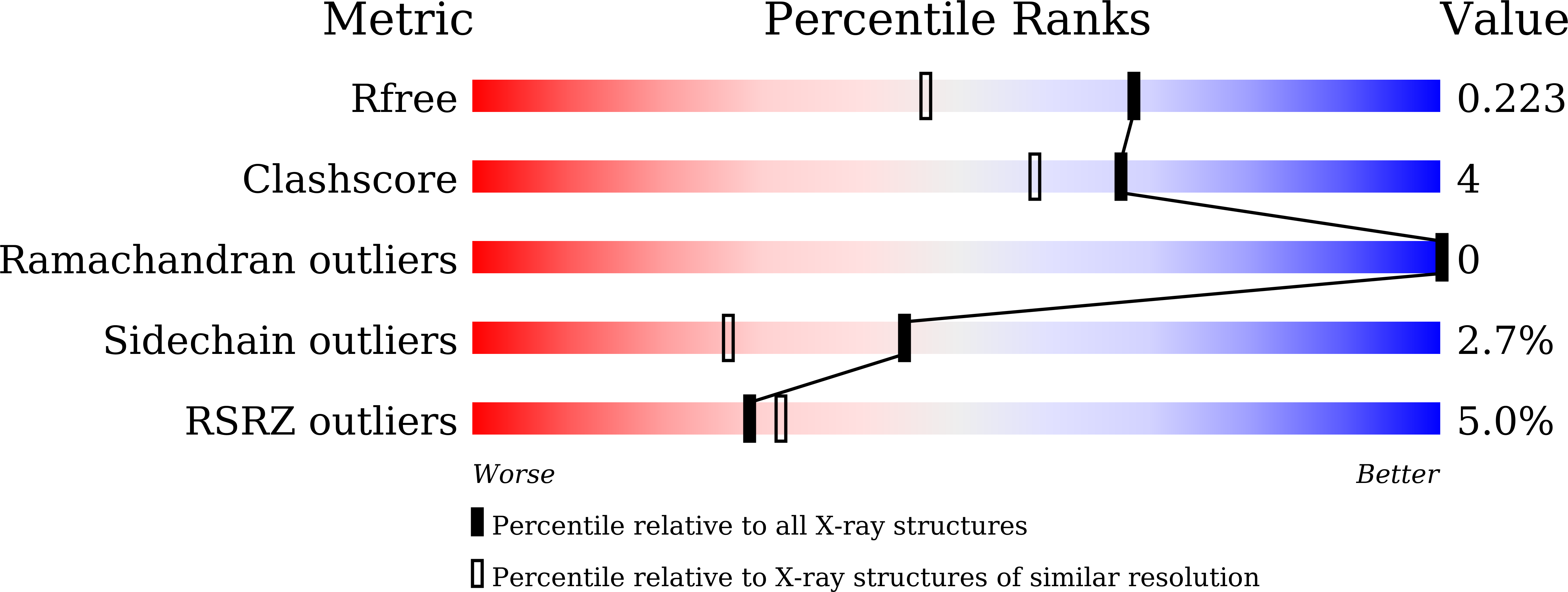
    5B68

  • PubMed Abstract: 

    Amylomaltase is an essential enzyme in maltose utilization and maltodextrin metabolism, and it has been industrially used for the production of cyclodextrin and modification of starch. We determined the crystal structure of amylomaltase from Corynebacterium glutamicum (CgAM) at a resolution of 1.7 Å. Although CgAM forms a dimer without NaCl, it exists as a monomer in physiological concentration of NaCl. CgAM is composed of N- and C-terminal domains, which can be further divided into two and four subdomains, respectively. It exhibits a unique structural feature at the functionally unknown N-domain and also shows two striking differences at the C-domain compared to other amylomaltases. These differences at extended edge of the substrate-binding site might affect substrate specificity for large cyclodextrin formation. The bis-tris methane and sulfate molecules bound at the substrate-binding site of our current structure mimic the binding of the hydroxyl groups of glucose bound at subsites -1 and -2, respectively.


  • Organizational Affiliation

    Structural and Molecular Biology Laboratory, School of Life Sciences and Biotechnology, Kyungpook National University , Daehak-ro 80, Buk-ku, Daegu 702-701, Republic of Korea.


Macromolecules
Find similar proteins by:  (by identity cutoff)  |  3D Structure
Entity ID: 1
MoleculeChains Sequence LengthOrganismDetailsImage
4-alpha-glucanotransferase714Corynebacterium glutamicumMutation(s): 0 
EC: 2.4.1.25
UniProt
Find proteins for Q8NNA7 (Corynebacterium glutamicum (strain ATCC 13032 / DSM 20300 / BCRC 11384 / JCM 1318 / LMG 3730 / NCIMB 10025))
Explore Q8NNA7 
Go to UniProtKB:  Q8NNA7
Entity Groups  
Sequence Clusters30% Identity50% Identity70% Identity90% Identity95% Identity100% Identity
UniProt GroupQ8NNA7
Sequence Annotations
Expand
  • Reference Sequence
Small Molecules
Ligands 3 Unique
IDChains Name / Formula / InChI Key2D Diagram3D Interactions
BTB
Query on BTB

Download Ideal Coordinates CCD File 
B [auth A]2-[BIS-(2-HYDROXY-ETHYL)-AMINO]-2-HYDROXYMETHYL-PROPANE-1,3-DIOL
C8 H19 N O5
OWMVSZAMULFTJU-UHFFFAOYSA-N
SO4
Query on SO4

Download Ideal Coordinates CCD File 
E [auth A]
F [auth A]
G [auth A]
H [auth A]
I [auth A]
E [auth A],
F [auth A],
G [auth A],
H [auth A],
I [auth A],
J [auth A],
K [auth A],
L [auth A],
M [auth A]
SULFATE ION
O4 S
QAOWNCQODCNURD-UHFFFAOYSA-L
GOL
Query on GOL

Download Ideal Coordinates CCD File 
C [auth A],
D [auth A]
GLYCEROL
C3 H8 O3
PEDCQBHIVMGVHV-UHFFFAOYSA-N
Experimental Data & Validation

Experimental Data

  • Method: X-RAY DIFFRACTION
  • Resolution: 1.70 Å
  • R-Value Free: 0.222 
  • R-Value Work: 0.181 
  • R-Value Observed: 0.199 
  • Space Group: P 21 21 21
Unit Cell:
Length ( Å )Angle ( ˚ )
a = 73.044α = 90
b = 83.251β = 90
c = 124.821γ = 90
Software Package:
Software NamePurpose
HKL-2000data collection
HKL-2000data scaling
REFMACrefinement
PDB_EXTRACTdata extraction
HKL-2000data reduction
MOLREPphasing

Structure Validation

View Full Validation Report


Entry History 

Deposition Data

Revision History  (Full details and data files)

  • Version 1.0: 2016-08-03
    Type: Initial release
  • Version 1.1: 2023-11-08
    Changes: Data collection, Database references, Derived calculations, Refinement description