Structural Insights into l-Tryptophan Dehydrogenase from a Photoautotrophic Cyanobacterium, Nostoc punctiforme.
Wakamatsu, T., Sakuraba, H., Kitamura, M., Hakumai, Y., Fukui, K., Ohnishi, K., Ashiuchi, M., Ohshima, T.(2017) Appl Environ Microbiol 83
- PubMed: 27815281
- DOI: https://doi.org/10.1128/AEM.02710-16
- Primary Citation of Related Structures:
5B37 - PubMed Abstract:
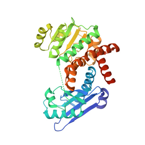
l-Tryptophan dehydrogenase from Nostoc punctiforme NIES-2108 (NpTrpDH), despite exhibiting high amino acid sequence identity (>30%)/homology (>50%) with NAD(P) + -dependent l-Glu/l-Leu/l-Phe/l-Val dehydrogenases, exclusively catalyzes reversible oxidative deamination of l-Trp to 3-indolepyruvate in the presence of NAD + Here, we determined the crystal structure of the apo form of NpTrpDH. The structure of the NpTrpDH monomer, which exhibited high similarity to that of l-Glu/l-Leu/l-Phe dehydrogenases, consisted of a substrate-binding domain (domain I, residues 3 to 133 and 328 to 343) and an NAD + /NADH-binding domain (domain II, residues 142 to 327) separated by a deep cleft. The apo-NpTrpDH existed in an open conformation, where domains I and II were apart from each other. The subunits dimerized themselves mainly through interactions between amino acid residues around the β-1 strand of each subunit, as was observed in the case of l-Phe dehydrogenase. The binding site for the substrate l-Trp was predicted by a molecular docking simulation and validated by site-directed mutagenesis. Several hydrophobic residues, which were located in the active site of NpTrpDH and possibly interacted with the side chain of the substrate l-Trp, were arranged similarly to that found in l-Leu/l-Phe dehydrogenases but fairly different from that of an l-Glu dehydrogenase. Our crystal structure revealed that Met-40, Ala-69, Ile-74, Ile-110, Leu-288, Ile-289, and Tyr-292 formed a hydrophobic cluster around the active site. The results of the site-directed mutagenesis experiments suggested that the hydrophobic cluster plays critical roles in protein folding, l-Trp recognition, and catalysis. Our results provide critical information for further characterization and engineering of this enzyme.
Organizational Affiliation:
Agricultural Science, Graduate School of Integrated Arts and Sciences, Kochi University, Kochi, Japan t-wakamatsu@kochi-u.ac.jp.