5B2N

Crystal structure of the light-driven chloride ion-pumping rhodopsin, ClP, from Nonlabens marinus


Experimental Data Snapshot

  • Method: X-RAY DIFFRACTION
  • Resolution: 1.58 Å
  • R-Value Free: 0.198 
  • R-Value Work: 0.176 
  • R-Value Observed: 0.177 

wwPDB Validation   3D Report Full Report


Ligand Structure Quality Assessment 


This is version 1.3 of the entry. See complete history


Literature

Structural Mechanism for Light-driven Transport by a New Type of Chloride Ion Pump, Nonlabens marinus Rhodopsin-3

Hosaka, T.Yoshizawa, S.Nakajima, Y.Ohsawa, N.Hato, M.DeLong, E.F.Kogure, K.Yokoyama, S.Kimura-Someya, T.Iwasaki, W.Shirouzu, M.

(2016) J Biol Chem 291: 17488-17495

  • DOI: https://doi.org/10.1074/jbc.M116.728220
  • Primary Citation of Related Structures:  
    5B2N

  • PubMed Abstract: 

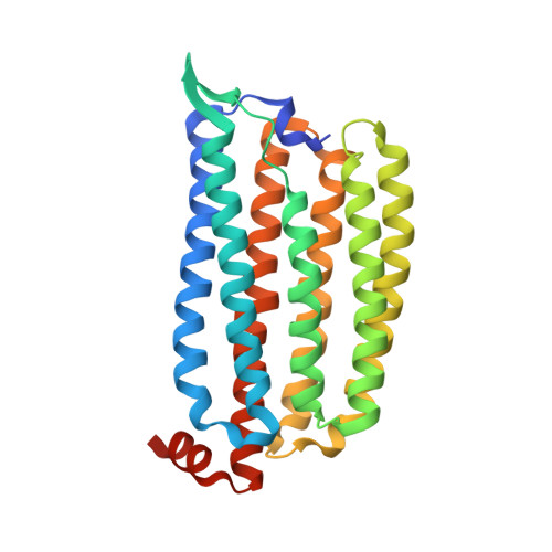
    The light-driven inward chloride ion-pumping rhodopsin Nonlabens marinus rhodopsin-3 (NM-R3), from a marine flavobacterium, belongs to a phylogenetic lineage distinct from the halorhodopsins known as archaeal inward chloride ion-pumping rhodopsins. NM-R3 and halorhodopsin have distinct motif sequences that are important for chloride ion binding and transport. In this study, we present the crystal structure of a new type of light-driven chloride ion pump, NM-R3, at 1.58 Å resolution. The structure revealed the chloride ion translocation pathway and showed that a single chloride ion resides near the Schiff base. The overall structure, chloride ion-binding site, and translocation pathway of NM-R3 are different from those of halorhodopsin. Unexpectedly, this NM-R3 structure is similar to the crystal structure of the light-driven outward sodium ion pump, Krokinobacter eikastus rhodopsin 2. Structural and mutational analyses of NM-R3 revealed that most of the important amino acid residues for chloride ion pumping exist in the ion influx region, located on the extracellular side of NM-R3. In contrast, on the opposite side, the cytoplasmic regions of K. eikastus rhodopsin 2 were reportedly important for sodium ion pumping. These results provide new insight into ion selection mechanisms in ion pumping rhodopsins, in which the ion influx regions of both the inward and outward pumps are important for their ion selectivities.


  • Organizational Affiliation

    From the Division of Structural and Synthetic Biology, RIKEN Center for Life Science Technologies, and.


Macromolecules
Find similar proteins by:  (by identity cutoff)  |  3D Structure
Entity ID: 1
MoleculeChains Sequence LengthOrganismDetailsImage
Chloride pumping rhodopsin279Nonlabens marinus S1-08Mutation(s): 0 
Gene Names: ClRNMS_1267
Membrane Entity: Yes 
UniProt
Find proteins for W8VZW3 (Nonlabens marinus S1-08)
Explore W8VZW3 
Go to UniProtKB:  W8VZW3
Entity Groups  
Sequence Clusters30% Identity50% Identity70% Identity90% Identity95% Identity100% Identity
UniProt GroupW8VZW3
Sequence Annotations
Expand
  • Reference Sequence
Small Molecules
Ligands 6 Unique
IDChains Name / Formula / InChI Key2D Diagram3D Interactions
RET
Query on RET

Download Ideal Coordinates CCD File 
B [auth A]RETINAL
C20 H28 O
NCYCYZXNIZJOKI-OVSJKPMPSA-N
D12
Query on D12

Download Ideal Coordinates CCD File 
J [auth A],
K [auth A],
L [auth A]
DODECANE
C12 H26
SNRUBQQJIBEYMU-UHFFFAOYSA-N
D10
Query on D10

Download Ideal Coordinates CCD File 
M [auth A],
N [auth A],
O [auth A]
DECANE
C10 H22
DIOQZVSQGTUSAI-UHFFFAOYSA-N
OCT
Query on OCT

Download Ideal Coordinates CCD File 
F [auth A],
G [auth A],
H [auth A],
I [auth A]
N-OCTANE
C8 H18
TVMXDCGIABBOFY-UHFFFAOYSA-N
HEX
Query on HEX

Download Ideal Coordinates CCD File 
D [auth A],
E [auth A]
HEXANE
C6 H14
VLKZOEOYAKHREP-UHFFFAOYSA-N
CL
Query on CL

Download Ideal Coordinates CCD File 
C [auth A]CHLORIDE ION
Cl
VEXZGXHMUGYJMC-UHFFFAOYSA-M
Experimental Data & Validation

Experimental Data

  • Method: X-RAY DIFFRACTION
  • Resolution: 1.58 Å
  • R-Value Free: 0.198 
  • R-Value Work: 0.176 
  • R-Value Observed: 0.177 
  • Space Group: C 1 2 1
Unit Cell:
Length ( Å )Angle ( ˚ )
a = 101.58α = 90
b = 49.79β = 130.4
c = 75.32γ = 90
Software Package:
Software NamePurpose
XSCALEdata scaling
PHASERphasing
PHENIXrefinement
PDB_EXTRACTdata extraction
XDSdata reduction

Structure Validation

View Full Validation Report


Ligand Structure Quality Assessment 


Entry History 

Deposition Data

Revision History  (Full details and data files)

  • Version 1.0: 2016-07-13
    Type: Initial release
  • Version 1.1: 2016-09-07
    Changes: Database references
  • Version 1.2: 2020-02-26
    Changes: Data collection, Database references, Derived calculations
  • Version 1.3: 2023-11-08
    Changes: Data collection, Database references, Refinement description