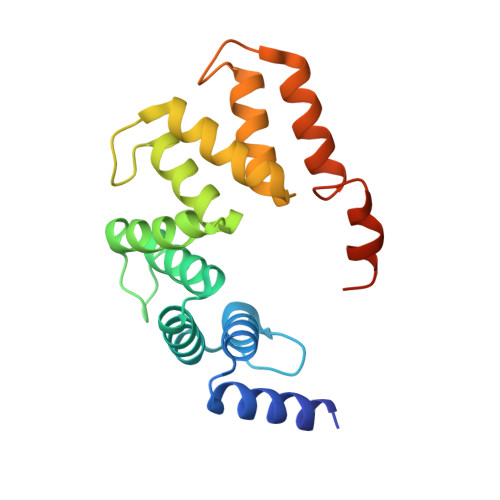
5B26

Crystal structure of mouse SEL1L

  • Classification: SIGNALING PROTEIN
  • Organism(s): Mus musculus
  • Expression System: Escherichia coli BL21(DE3)
  • Mutation(s): No 

  • Deposited: 2016-01-09 Released: 2016-04-27 
  • Deposition Author(s): Jeong, H., Lee, C.
  • Funding Organization(s): Ministry of Health & Welfare, Ulsan National Institute of Science and Technology, National Research Foundation of Korea

Experimental Data Snapshot

  • Method: X-RAY DIFFRACTION
  • Resolution: 2.60 Å
  • R-Value Free: 0.282 
  • R-Value Work: 0.207 
  • R-Value Observed: 0.210 

wwPDB Validation   3D Report Full Report


This is version 1.1 of the entry. See complete history


Literature

Crystal structure of SEL1L: Insight into the roles of SLR motifs in ERAD pathway

Jeong, H.Sim, H.J.Song, E.K.Lee, H.Ha, S.C.Jun, Y.Park, T.J.Lee, C.

(2016) Sci Rep 6: 20261-20261

  • DOI: https://doi.org/10.1038/srep20261
  • Primary Citation of Related Structures:  
    5B26

  • PubMed Abstract: 

    Terminally misfolded proteins are selectively recognized and cleared by the endoplasmic reticulum-associated degradation (ERAD) pathway. SEL1L, a component of the ERAD machinery, plays an important role in selecting and transporting ERAD substrates for degradation. We have determined the crystal structure of the mouse SEL1L central domain comprising five Sel1-Like Repeats (SLR motifs 5 to 9; hereafter called SEL1L(cent)). Strikingly, SEL1L(cent) forms a homodimer with two-fold symmetry in a head-to-tail manner. Particularly, the SLR motif 9 plays an important role in dimer formation by adopting a domain-swapped structure and providing an extensive dimeric interface. We identified that the full-length SEL1L forms a self-oligomer through the SEL1L(cent) domain in mammalian cells. Furthermore, we discovered that the SLR-C, comprising SLR motifs 10 and 11, of SEL1L directly interacts with the N-terminus luminal loops of HRD1. Therefore, we propose that certain SLR motifs of SEL1L play a unique role in membrane bound ERAD machinery.


  • Organizational Affiliation

    Department of Biological Sciences, School of Life Sciences, Ulsan National Institute of Science and Technology, 50 UNIST-gil, Ulsan 44919, Republic of Korea.


Macromolecules
Find similar proteins by:  (by identity cutoff)  |  3D Structure
Entity ID: 1
MoleculeChains Sequence LengthOrganismDetailsImage
Protein sel-1 homolog 1
A, B, C, D
186Mus musculusMutation(s): 0 
Gene Names: Sel1lSel1h
UniProt & NIH Common Fund Data Resources
Find proteins for Q9Z2G6 (Mus musculus)
Explore Q9Z2G6 
Go to UniProtKB:  Q9Z2G6
IMPC:  MGI:1329016
Entity Groups  
Sequence Clusters30% Identity50% Identity70% Identity90% Identity95% Identity100% Identity
UniProt GroupQ9Z2G6
Sequence Annotations
Expand
  • Reference Sequence
Experimental Data & Validation

Experimental Data

  • Method: X-RAY DIFFRACTION
  • Resolution: 2.60 Å
  • R-Value Free: 0.282 
  • R-Value Work: 0.207 
  • R-Value Observed: 0.210 
  • Space Group: P 1 21 1
Unit Cell:
Length ( Å )Angle ( ˚ )
a = 29.128α = 90
b = 110.523β = 90.61
c = 109.81γ = 90
Software Package:
Software NamePurpose
PHENIXrefinement
HKL-2000data processing
PHENIXphasing
Cootmodel building

Structure Validation

View Full Validation Report


Entry History & Funding Information

Deposition Data


Funding OrganizationLocationGrant Number
Ministry of Health & WelfareKorea, Republic OfHI12C1744
Ulsan National Institute of Science and TechnologyKorea, Republic Of1.150024.01
National Research Foundation of KoreaKorea, Republic OfNRF-2011-0024241
National Research Foundation of KoreaKorea, Republic Of2012R1A1A1011604
National Research Foundation of KoreaKorea, Republic OfNRF-2014H1A2A1020322

Revision History  (Full details and data files)

  • Version 1.0: 2016-04-27
    Type: Initial release
  • Version 1.1: 2024-03-20
    Changes: Data collection, Database references, Derived calculations