


Crystal structure of Bacillus stearothermophilus Farnesyl pyrophosphate synthase
Makabe, K., Kijima, T.To be published.
Experimental Data Snapshot
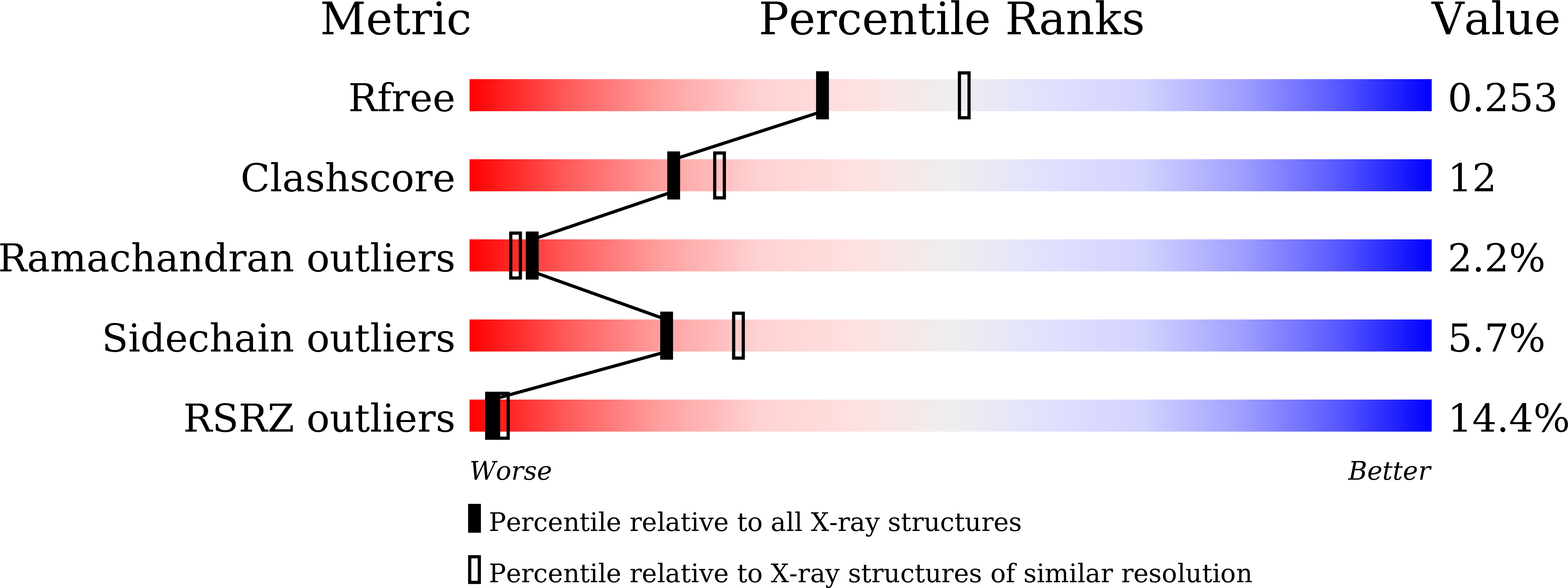
wwPDB Validation 3D Report Full Report
Entity ID: 1 | |||||
|---|---|---|---|---|---|
| Molecule | Chains | Sequence Length | Organism | Details | Image |
| Farnesyl diphosphate synthase | 297 | Geobacillus stearothermophilus | Mutation(s): 0 EC: 2.5.1.10 | 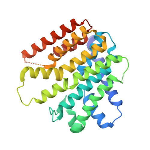 | |
UniProt | |||||
Find proteins for Q08291 (Geobacillus stearothermophilus) Explore Q08291 Go to UniProtKB: Q08291 | |||||
Entity Groups | |||||
| Sequence Clusters | 30% Identity50% Identity70% Identity90% Identity95% Identity100% Identity | ||||
| UniProt Group | Q08291 | ||||
Sequence AnnotationsExpand | |||||
| |||||
| Length ( Å ) | Angle ( ˚ ) |
|---|---|
| a = 49.015 | α = 90 |
| b = 95.156 | β = 90 |
| c = 103.349 | γ = 90 |
| Software Name | Purpose |
|---|---|
| REFMAC | refinement |
| Coot | model building |
RCSB PDB (citation) is hosted by
RCSB PDB is a member of the

