5AYH

Structure of the entire dynein stalk region


Experimental Data Snapshot

  • Method: X-RAY DIFFRACTION
  • Resolution: 3.01 Å
  • R-Value Free: 0.295 
  • R-Value Work: 0.255 
  • R-Value Observed: 0.259 

wwPDB Validation   3D Report Full Report


This is version 1.3 of the entry. See complete history

Re-refinement Note

This entry reflects an alternative modeling of the original data in: 3WUQ


Literature

Structural Change in the Dynein Stalk Region Associated with Two Different Affinities for the Microtubule

Nishikawa, Y.Inatomi, M.Iwasaki, H.Kurisu, G.

(2016) J Mol Biol 428: 1886-1896

  • DOI: https://doi.org/10.1016/j.jmb.2015.11.008
  • Primary Citation of Related Structures:  
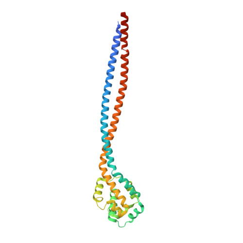
    5AYH

  • PubMed Abstract: 

    Dynein is a large microtubule-based motor complex that requires tight coupling of intra-molecular ATP hydrolysis with the generation of mechanical force and track-binding activity. However, the microtubule-binding domain is structurally separated by about 15nm from the nucleotide-binding sites by a coiled-coil stalk. Thus, long-range two-way communication is necessary for coordination between the catalytic cycle of ATP hydrolysis and dynein's track-binding affinities. To investigate the structural changes that occur in the dynein stalk region to produce two different microtubule affinities, here we improve the resolution limit of the previously reported structure of the entire stalk region and we investigate structural changes in the dynein stalk and strut/buttress regions by comparing currently available X-ray structures. In the light of recent crystal structures, the basis of the transition from the low-affinity to the high-affinity coiled-coil registry is discussed. A concerted movement model previously reported by Carter and Vale is modified more specifically, and we proposed it as the open zipper model.


  • Organizational Affiliation

    Institute for Protein Research, Osaka University, Suita, Osaka 565-0871, Japan.


Macromolecules
Find similar proteins by:  (by identity cutoff)  |  3D Structure
Entity ID: 1
MoleculeChains Sequence LengthOrganismDetailsImage
Cytoplasmic dynein 1 heavy chain 1276Mus musculusMutation(s): 0 
Gene Names: Dync1h1Dhc1Dnch1Dnchc1Dyhc
UniProt & NIH Common Fund Data Resources
Find proteins for Q9JHU4 (Mus musculus)
Explore Q9JHU4 
Go to UniProtKB:  Q9JHU4
IMPC:  MGI:103147
Entity Groups  
Sequence Clusters30% Identity50% Identity70% Identity90% Identity95% Identity100% Identity
UniProt GroupQ9JHU4
Sequence Annotations
Expand
  • Reference Sequence
Experimental Data & Validation

Experimental Data

  • Method: X-RAY DIFFRACTION
  • Resolution: 3.01 Å
  • R-Value Free: 0.295 
  • R-Value Work: 0.255 
  • R-Value Observed: 0.259 
  • Space Group: P 31 2 1
Unit Cell:
Length ( Å )Angle ( ˚ )
a = 103.325α = 90
b = 103.325β = 90
c = 69.709γ = 120
Software Package:
Software NamePurpose
PHENIXrefinement
HKL-2000data reduction
HKL-2000data scaling
PHASERphasing
Cootmodel building

Structure Validation

View Full Validation Report


Entry History & Funding Information

Deposition Data


Funding OrganizationLocationGrant Number
JSPSJapan26291014

Revision History  (Full details and data files)

  • Version 1.0: 2015-12-16
    Type: Initial release
  • Version 1.1: 2016-05-18
    Changes: Database references
  • Version 1.2: 2020-02-26
    Changes: Data collection, Database references, Derived calculations
  • Version 1.3: 2023-11-08
    Changes: Data collection, Database references, Refinement description