5AWT

Crystal structure of the SGIP1 mu homology domain in complex with an Eps15 fragment containing two DPF motifs (YDPFGGDPFKG)


Experimental Data Snapshot

  • Method: X-RAY DIFFRACTION
  • Resolution: 2.70 Å
  • R-Value Free: 0.234 
  • R-Value Work: 0.188 
  • R-Value Observed: 0.191 

wwPDB Validation   3D Report Full Report


This is version 1.2 of the entry. See complete history


Literature

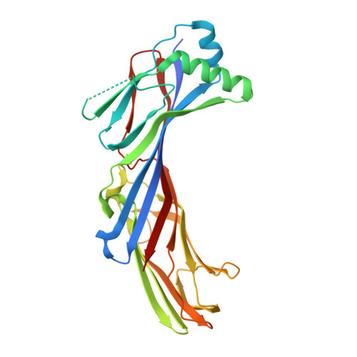
Structural basis for the recognition of two consecutive mutually interacting DPF motifs by the SGIP1 mu homology domain.

Shimada, A.Yamaguchi, A.Kohda, D.

(2016) Sci Rep 6: 19565-19565

  • DOI: https://doi.org/10.1038/srep19565
  • Primary Citation of Related Structures:  
    5AWR, 5AWS, 5AWT, 5AWU

  • PubMed Abstract: 

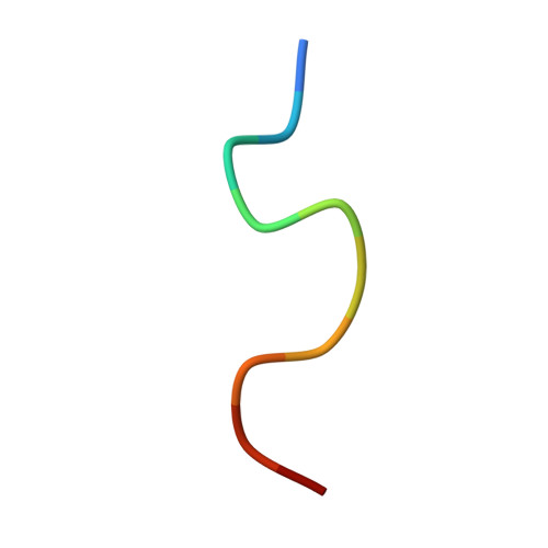
    FCHo1, FCHo2, and SGIP1 are key regulators of clathrin-mediated endocytosis. Their μ homology domains (μHDs) interact with the C-terminal region of an endocytic scaffold protein, Eps15, containing fifteen Asp-Pro-Phe (DPF) motifs. Here, we show that the high-affinity μHD-binding site in Eps15 is a region encompassing six consecutive DPF motifs, while the minimal μHD-binding unit is two consecutive DPF motifs. We present the crystal structures of the SGIP1 μHD in complex with peptides containing two DPF motifs. The peptides bind to a novel ligand-binding site of the μHD, which is distinct from those of other distantly related μHD-containing proteins. The two DPF motifs, which adopt three-dimensional structures stabilized by sequence-specific intramotif and intermotif interactions, are extensively recognized by the μHD and are both required for binding. Thus, consecutive and singly scattered DPF motifs play distinct roles in μHD binding.


  • Organizational Affiliation

    Division of Structural Biology, Medical Institute of Bioregulation, Kyushu University, 3-1-1 Maidashi, Higashi-ku, Fukuoka 812-8582, Japan.


Macromolecules
Find similar proteins by:  (by identity cutoff)  |  3D Structure
Entity ID: 1
MoleculeChains Sequence LengthOrganismDetailsImage
SH3-containing GRB2-like protein 3-interacting protein 1282Homo sapiensMutation(s): 0 
Gene Names: SGIP1
UniProt & NIH Common Fund Data Resources
Find proteins for Q9BQI5 (Homo sapiens)
Explore Q9BQI5 
Go to UniProtKB:  Q9BQI5
PHAROS:  Q9BQI5
GTEx:  ENSG00000118473 
Entity Groups  
Sequence Clusters30% Identity50% Identity70% Identity90% Identity95% Identity100% Identity
UniProt GroupQ9BQI5
Sequence Annotations
Expand
  • Reference Sequence

Find similar proteins by:  Sequence   |   3D Structure  

Entity ID: 2
MoleculeChains Sequence LengthOrganismDetailsImage
Epidermal growth factor receptor substrate 1511Homo sapiensMutation(s): 0 
UniProt & NIH Common Fund Data Resources
Find proteins for P42566 (Homo sapiens)
Explore P42566 
Go to UniProtKB:  P42566
PHAROS:  P42566
GTEx:  ENSG00000085832 
Entity Groups  
Sequence Clusters30% Identity50% Identity70% Identity90% Identity95% Identity100% Identity
UniProt GroupP42566
Sequence Annotations
Expand
  • Reference Sequence
Experimental Data & Validation

Experimental Data

  • Method: X-RAY DIFFRACTION
  • Resolution: 2.70 Å
  • R-Value Free: 0.234 
  • R-Value Work: 0.188 
  • R-Value Observed: 0.191 
  • Space Group: P 4 21 2
Unit Cell:
Length ( Å )Angle ( ˚ )
a = 107.716α = 90
b = 107.716β = 90
c = 79.978γ = 90
Software Package:
Software NamePurpose
PHENIXrefinement
HKL-2000data reduction
HKL-2000data scaling
PHENIXphasing

Structure Validation

View Full Validation Report


Entry History & Funding Information

Deposition Data


Funding OrganizationLocationGrant Number
MEXT/JSPSJapanKAKENHI 20687006
MEXT/JSPSJapanKAKENHI 24687014
MEXT/JSPSJapanKAKENHI 25121726

Revision History  (Full details and data files)

  • Version 1.0: 2016-07-06
    Type: Initial release
  • Version 1.1: 2020-02-26
    Changes: Data collection, Derived calculations
  • Version 1.2: 2023-11-08
    Changes: Data collection, Database references, Derived calculations, Refinement description