5AUI

Crystal structure of Ferredoxin from Thermosynechococcus elongatus


Experimental Data Snapshot

  • Method: X-RAY DIFFRACTION
  • Resolution: 1.50 Å
  • R-Value Free: 0.183 
  • R-Value Work: 0.159 

wwPDB Validation   3D Report Full Report


This is version 3.1 of the entry. See complete history


Literature

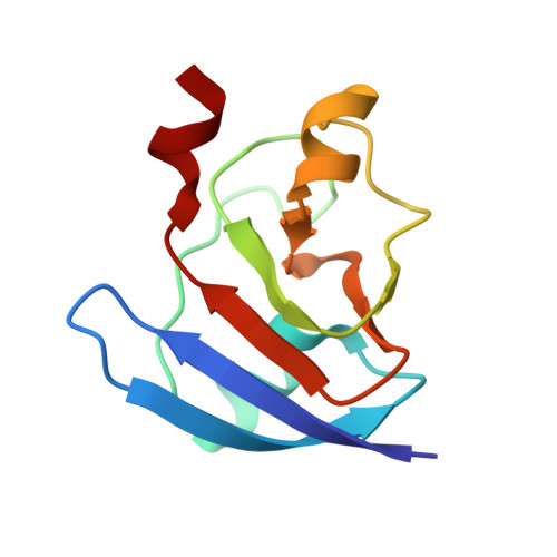
X-ray Structure and Nuclear Magnetic Resonance Analysis of the Interaction Sites of the Ga-Substituted Cyanobacterial Ferredoxin

Mutoh, R.Muraki, N.Shinmura, K.Kubota-Kawai, H.Lee, Y.H.Nowaczyk, M.M.Rogner, M.Hase, T.Ikegami, T.Kurisu, G.

(2015) Biochemistry 54: 6052-6061

  • DOI: https://doi.org/10.1021/acs.biochem.5b00601
  • Primary Citation of Related Structures:  
    5AUI, 5AUK

  • PubMed Abstract: 

    In chloroplasts, ferredoxin (Fd) is reduced by Photosystem I (PSI) and oxidized by Fd-NADP(+) reductase (FNR) that is involved in NADP(+) reduction. To understand the structural basis for the dynamics and efficiency of the electron transfer reaction via Fd, we complementary used X-ray crystallography and nuclear magnetic resonance (NMR) spectroscopy. In the NMR analysis of the formed electron transfer complex with Fd, the paramagnetic effect of the [2Fe-2S] cluster of Fd prevented us from detecting the NMR signals around the cluster. To solve this problem, the paramagnetic iron-sulfur cluster was replaced with a diamagnetic metal cluster. We determined the crystal structure of the Ga-substituted Fd (GaFd) from Synechocystis sp. PCC6803 at 1.62 Å resolution and verified its functional complementation using affinity chromatography. NMR analysis of the interaction sites on GaFd with PSI (molecular mass of ∼1 MDa) and FNR from Thermosynechococcus elongatus was achieved with high-field NMR spectroscopy. With reference to the interaction sites with FNR of Anabaena sp. PCC 7119 from the published crystal data, the interaction sites of Fd with FNR and PSI in solution can be classified into two types: (1) the core hydrophobic residues in the proximity of the metal center and (2) the hydrophilic residues surrounding the core. The former sites are shared in the Fd:FNR and Fd:PSI complex, while the latter ones are target-specific and not conserved on the residual level.


  • Organizational Affiliation

    Institute for Protein Research, Osaka University , 3-2 Yamadaoka, Suita, Osaka 565-0871, Japan.


Macromolecules
Find similar proteins by:  (by identity cutoff)  |  3D Structure
Entity ID: 1
MoleculeChains Sequence LengthOrganismDetailsImage
Ferredoxin-197Thermosynechococcus vestitus BP-1Mutation(s): 0 
Gene Names: petF1petFtsl1009
UniProt
Find proteins for P0A3C9 (Thermosynechococcus vestitus (strain NIES-2133 / IAM M-273 / BP-1))
Explore P0A3C9 
Go to UniProtKB:  P0A3C9
Entity Groups  
Sequence Clusters30% Identity50% Identity70% Identity90% Identity95% Identity100% Identity
UniProt GroupP0A3C9
Sequence Annotations
Expand
  • Reference Sequence
Experimental Data & Validation

Experimental Data

Unit Cell:
Length ( Å )Angle ( ˚ )
a = 56.441α = 90
b = 53.314β = 92.384
c = 32.278γ = 90
Software Package:
Software NamePurpose
REFMACrefinement
MOLREPphasing
Cootmodel building
HKL-2000data reduction
HKL-2000data scaling

Structure Validation

View Full Validation Report


Entry History & Funding Information

Deposition Data


Funding OrganizationLocationGrant Number
Japan Society for the Promotion of Science (JSPS)JapanGS016 (Cabinet Office of Japan)

Revision History  (Full details and data files)

  • Version 1.0: 2015-09-23
    Type: Initial release
  • Version 1.1: 2015-10-14
    Changes: Database references
  • Version 1.2: 2020-02-19
    Changes: Data collection, Database references, Derived calculations, Source and taxonomy
  • Version 2.0: 2021-08-18
    Changes: Atomic model, Data collection, Database references, Derived calculations, Non-polymer description, Structure summary
  • Version 3.0: 2022-02-09
    Type: Coordinate replacement
    Reason: Model completeness
    Changes: Advisory, Atomic model, Author supporting evidence, Data collection, Database references, Derived calculations, Non-polymer description, Other, Polymer sequence, Refinement description, Source and taxonomy, Structure summary
  • Version 3.1: 2023-11-08
    Changes: Data collection, Refinement description