5AQ1

Trypanosoma cruzi Glucose-6-phosphate Dehydrogenase in complex with G6P and NADPH


Experimental Data Snapshot

  • Method: X-RAY DIFFRACTION
  • Resolution: 2.65 Å
  • R-Value Free: 0.226 
  • R-Value Work: 0.200 
  • R-Value Observed: 0.201 

wwPDB Validation   3D Report Full Report


Ligand Structure Quality Assessment 


This is version 1.3 of the entry. See complete history


Literature

The Structure of a Trypanosoma Cruzi Glucose-6-Phosphate Dehydrogenase Reveals Differences from the Mammalian Enzyme.

Mercaldi, G.F.Dawson, A.Hunter, W.N.Cordeiro, A.T.

(2016) FEBS Lett 590: 2776

  • DOI: https://doi.org/10.1002/1873-3468.12276
  • Primary Citation of Related Structures:  
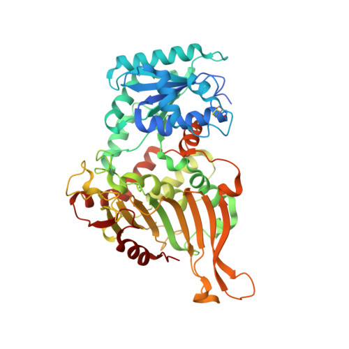
    5AQ1

  • PubMed Abstract: 

    The enzyme glucose-6-phosphate dehydrogenase from Trypanosoma cruzi (TcG6PDH) catalyses the first step of the pentose phosphate pathway (PPP) and is considered a promising target for the discovery of a new drug against Chagas diseases. In the present work, we describe the crystal structure of TcG6PDH obtained in a ternary complex with the substrate β-d-glucose-6-phosphate (G6P) and the reduced 'catalytic' cofactor NADPH, which reveals the molecular basis of substrate and cofactor recognition. A comparison with the homologous human protein sheds light on differences in the cofactor-binding site that might be explored towards the design of new NADP(+) competitive inhibitors targeting the parasite enzyme.


  • Organizational Affiliation

    Brazilian Biosciences National Laboratory, Center of Research in Energy and Materials, Campinas, Brazil.


Macromolecules
Find similar proteins by:  (by identity cutoff)  |  3D Structure
Entity ID: 1
MoleculeChains Sequence LengthOrganismDetailsImage
GLUCOSE-6-PHOSPHATE DEHYDROGENASE
A, B, C
509Trypanosoma cruziMutation(s): 0 
EC: 1.1.1.49
UniProt
Find proteins for Q4E0B2 (Trypanosoma cruzi (strain CL Brener))
Explore Q4E0B2 
Go to UniProtKB:  Q4E0B2
Entity Groups  
Sequence Clusters30% Identity50% Identity70% Identity90% Identity95% Identity100% Identity
UniProt GroupQ4E0B2
Sequence Annotations
Expand
  • Reference Sequence
Experimental Data & Validation

Experimental Data

  • Method: X-RAY DIFFRACTION
  • Resolution: 2.65 Å
  • R-Value Free: 0.226 
  • R-Value Work: 0.200 
  • R-Value Observed: 0.201 
  • Space Group: I 41 2 2
Unit Cell:
Length ( Å )Angle ( ˚ )
a = 154.946α = 90
b = 154.946β = 90
c = 348.358γ = 90
Software Package:
Software NamePurpose
REFMACrefinement
XDSdata reduction
Aimlessdata scaling
PHASERphasing

Structure Validation

View Full Validation Report


Ligand Structure Quality Assessment 


Entry History 

Deposition Data

Revision History  (Full details and data files)

  • Version 1.0: 2016-07-20
    Type: Initial release
  • Version 1.1: 2016-09-28
    Changes: Database references
  • Version 1.2: 2020-07-29
    Type: Remediation
    Reason: Carbohydrate remediation
    Changes: Data collection, Derived calculations, Other, Structure summary
  • Version 1.3: 2024-01-10
    Changes: Data collection, Database references, Refinement description, Structure summary