5AN1

Crystallographic structure of the Glutathione S-Transferase from Litopenaeus vannamei complexed with Glutathione


Experimental Data Snapshot

  • Method: X-RAY DIFFRACTION
  • Resolution: 2.00 Å
  • R-Value Free: 0.262 
  • R-Value Work: 0.214 
  • R-Value Observed: 0.215 

wwPDB Validation   3D Report Full Report


Ligand Structure Quality Assessment 


This is version 1.3 of the entry. See complete history


Literature

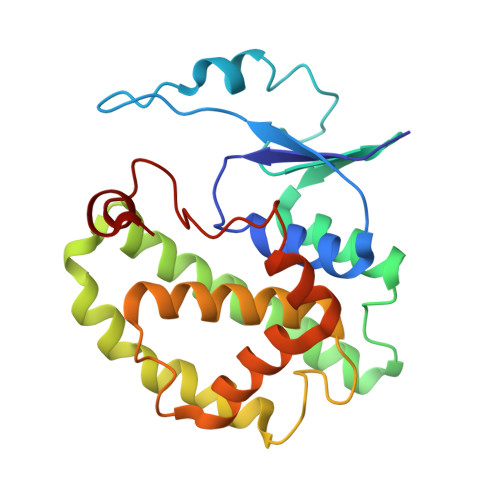
Crystal structure of a class-mu glutathione S-transferase from whiteleg shrimp Litopenaeus vannamei: structural changes in the xenobiotic binding H-site may alter the spectra of molecules bound.

Juarez-Martinez, A.B.Sotelo-Mundo, R.R.Rudino-Pinera, E.

(2017) J Biochem Mol Toxicol 31

  • DOI: https://doi.org/10.1002/jbt.21838
  • Primary Citation of Related Structures:  
    5AN1

  • PubMed Abstract: 

    Glutathione S-transferases (GSTs) are dimeric proteins that play a key role in phase II cellular detoxification. Here, the first crystal structure of a GST class-mu from marine crustacean shrimp Litopenaeus vannamei is reported at a resolution of 2.0 Å. The coordinates reported here have the lowest sequence identity with previously reported GSTs class-mu deposited at the Protein Data Bank (PDB), although they have subtle conformational differences. One key feature of GST class-mu from L. vannamei is the active site crevice markedly reduced when it is compared with other GSTs class-mu. This finding together with the chemical change of residues into the cavity (F112 and Y210) points to a particular specialization in which smallest xenobiotics with nonstandard chemical characteristics can be bound to the H-site. This suggests that marine organisms have evolved structural strategies to provide efficient selectivity toward xenobiotics to be disposed of by the phase II detoxification process.


  • Organizational Affiliation

    Departamento de Medicina Molecular y Bioprocesos, Instituto de Biotecnología, Universidad Nacional Autónoma de México, Colonia Chamilpa, 62210, Cuernavaca, Morelos, México.


Macromolecules
Find similar proteins by:  (by identity cutoff)  |  3D Structure
Entity ID: 1
MoleculeChains Sequence LengthOrganismDetailsImage
GLUTATHIONE S-TRANSFERASE
A, B, C, D, E
A, B, C, D, E, F, G, H
219Penaeus vannameiMutation(s): 0 
EC: 2.5.1.18
UniProt
Find proteins for Q49SB0 (Penaeus vannamei)
Explore Q49SB0 
Go to UniProtKB:  Q49SB0
Entity Groups  
Sequence Clusters30% Identity50% Identity70% Identity90% Identity95% Identity100% Identity
UniProt GroupQ49SB0
Sequence Annotations
Expand
  • Reference Sequence
Experimental Data & Validation

Experimental Data

  • Method: X-RAY DIFFRACTION
  • Resolution: 2.00 Å
  • R-Value Free: 0.262 
  • R-Value Work: 0.214 
  • R-Value Observed: 0.215 
  • Space Group: P 1 21 1
Unit Cell:
Length ( Å )Angle ( ˚ )
a = 62.025α = 90
b = 101.943β = 90.07
c = 166.595γ = 90
Software Package:
Software NamePurpose
PHENIXrefinement
XDSdata reduction
XSCALEdata scaling
PHASERphasing

Structure Validation

View Full Validation Report


Ligand Structure Quality Assessment 


Entry History 

Deposition Data

Revision History  (Full details and data files)

  • Version 1.0: 2016-10-12
    Type: Initial release
  • Version 1.1: 2016-10-19
    Changes: Database references
  • Version 1.2: 2017-03-22
    Changes: Database references
  • Version 1.3: 2024-01-10
    Changes: Data collection, Database references, Derived calculations, Other, Refinement description