5AFZ

Thrombin in complex with (2R)-2-(benzylsulfonylamino)-N-(2-((4- carbamimidoylphenyl)methylamino)-2-oxo-propyl)-3-phenyl-propanamide


Experimental Data Snapshot

  • Method: X-RAY DIFFRACTION
  • Resolution: 1.53 Å
  • R-Value Free: 0.174 
  • R-Value Work: 0.145 
  • R-Value Observed: 0.146 

wwPDB Validation   3D Report Full Report


Ligand Structure Quality Assessment 


This is version 2.1 of the entry. See complete history


Literature

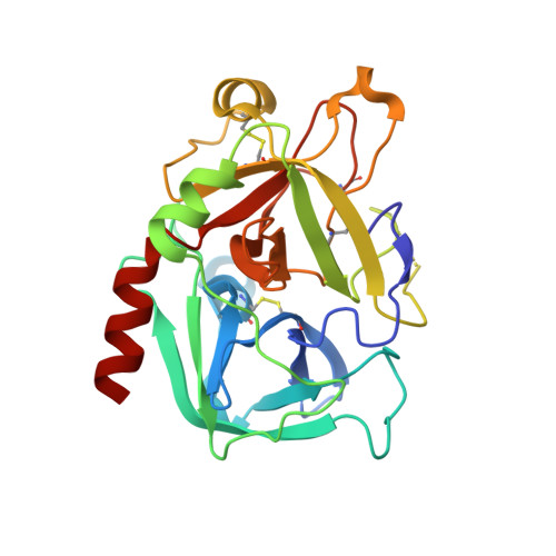
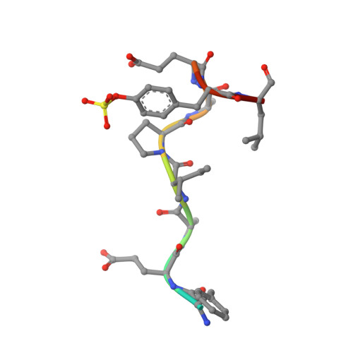
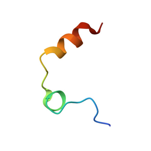
Fragments Can Bind Either More Enthalpy or Entropy-Driven: Crystal Structures and Residual Hydration Pattern Suggest Why.

Ruehmann, E.Betz, M.Heine, A.Klebe, G.

(2015) J Med Chem 58: 6960

  • DOI: https://doi.org/10.1021/acs.jmedchem.5b00812
  • Primary Citation of Related Structures:  
    4UD9, 4UDW, 4UE7, 4UEH, 5AF9, 5AFY, 5AFZ, 5AHG

  • PubMed Abstract: 

    In lead optimization, small, enthalpically advantaged fragments have been suggested to be superior, as an entropic component will be added inevitably during late-stage optimization. Determination of thermodynamic signatures of weak-binding fragments is essential to support the decision-making process, to decide which fragment to take to further optimization. High-resolution crystal structures of six fragments binding to the S1 pocket of thrombin were determined and analyzed with respect to their thermodynamic profile. The two most potent fragments exhibiting an amidine-type scaffold are not the most enthalpic binders; instead a chloro-thiophene fragment binds more enthalpically. Two chemically very similar chloro-aromatic fragments differ strongly in their potency (430 μM vs 10 mM); their binding modes are related, but the surrounding residual water network differs. The more potent one recruits a water molecule and involves Glu192 in binding, thus succeeding in firmly capping the S1 pocket. Fragments exhibiting a rather perfect solvation pattern in their binding mode also experience the highest potency.


  • Organizational Affiliation

    Institute of Pharmaceutical Chemistry, Philipps-University Marburg , Marbacher Weg 6, 35037 Marburg, Germany.


Macromolecules
Find similar proteins by:  (by identity cutoff)  |  3D Structure
Entity ID: 1
MoleculeChains Sequence LengthOrganismDetailsImage
ProthrombinA [auth H]258Homo sapiensMutation(s): 0 
EC: 3.4.21.5
UniProt & NIH Common Fund Data Resources
Find proteins for P00734 (Homo sapiens)
Explore P00734 
Go to UniProtKB:  P00734
PHAROS:  P00734
GTEx:  ENSG00000180210 
Entity Groups  
Sequence Clusters30% Identity50% Identity70% Identity90% Identity95% Identity100% Identity
UniProt GroupP00734
Sequence Annotations
Expand
  • Reference Sequence

Find similar proteins by:  Sequence   |   3D Structure  

Entity ID: 2
MoleculeChains Sequence LengthOrganismDetailsImage
Hirudin-2B [auth I]12Hirudo medicinalisMutation(s): 0 
UniProt
Find proteins for P09945 (Hirudo medicinalis)
Explore P09945 
Go to UniProtKB:  P09945
Entity Groups  
Sequence Clusters30% Identity50% Identity70% Identity90% Identity95% Identity100% Identity
UniProt GroupP09945
Sequence Annotations
Expand
  • Reference Sequence
Find similar proteins by:  (by identity cutoff)  |  3D Structure
Entity ID: 3
MoleculeChains Sequence LengthOrganismDetailsImage
ProthrombinC [auth L]29Homo sapiensMutation(s): 0 
EC: 3.4.21.5
UniProt & NIH Common Fund Data Resources
Find proteins for P00734 (Homo sapiens)
Explore P00734 
Go to UniProtKB:  P00734
PHAROS:  P00734
GTEx:  ENSG00000180210 
Entity Groups  
Sequence Clusters30% Identity50% Identity70% Identity90% Identity95% Identity100% Identity
UniProt GroupP00734
Sequence Annotations
Expand
  • Reference Sequence
Small Molecules
Modified Residues  1 Unique
IDChains TypeFormula2D DiagramParent
TYS
Query on TYS
B [auth I]L-PEPTIDE LINKINGC9 H11 N O6 STYR
Binding Affinity Annotations 
IDSourceBinding Affinity
UET BindingDB:  5AFZ Ki: 140 (nM) from 1 assay(s)
Kd: 160 (nM) from 1 assay(s)
Binding MOAD:  5AFZ Kd: 160 (nM) from 1 assay(s)
Experimental Data & Validation

Experimental Data

  • Method: X-RAY DIFFRACTION
  • Resolution: 1.53 Å
  • R-Value Free: 0.174 
  • R-Value Work: 0.145 
  • R-Value Observed: 0.146 
  • Space Group: C 1 2 1
Unit Cell:
Length ( Å )Angle ( ˚ )
a = 70.308α = 90
b = 71.643β = 100.53
c = 72.354γ = 90
Software Package:
Software NamePurpose
PHENIXrefinement
XDSdata reduction
XDSdata scaling
PHASERphasing

Structure Validation

View Full Validation Report


Ligand Structure Quality Assessment 


Entry History 

Deposition Data

Revision History  (Full details and data files)

  • Version 1.0: 2015-08-26
    Type: Initial release
  • Version 1.1: 2015-09-23
    Changes: Database references
  • Version 1.2: 2020-07-29
    Type: Remediation
    Reason: Carbohydrate remediation
    Changes: Data collection, Derived calculations, Other, Structure summary
  • Version 2.0: 2022-12-07
    Changes: Advisory, Atomic model, Data collection, Database references, Derived calculations, Polymer sequence, Source and taxonomy, Structure summary
  • Version 2.1: 2024-01-31
    Changes: Data collection, Refinement description