5ADH

INTERDOMAIN MOTION IN LIVER ALCOHOL DEHYDROGENASE. STRUCTURAL AND ENERGETIC ANALYSIS OF THE HINGE BENDING MODE


Experimental Data Snapshot

  • Method: X-RAY DIFFRACTION
  • Resolution: 2.90 Å

wwPDB Validation   3D Report Full Report


This is version 2.0 of the entry. See complete history


Literature

Interdomain motion in liver alcohol dehydrogenase. Structural and energetic analysis of the hinge bending mode.

Colonna-Cesari, F.Perahia, D.Karplus, M.Eklund, H.Braden, C.I.Tapia, O.

(1986) J Biol Chem 261: 15273-15280

  • Primary Citation of Related Structures:  
    5ADH, 8ADH

  • PubMed Abstract: 

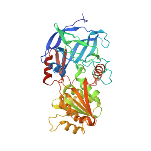
    A study of the hinge bending mode in the enzyme liver alcohol dehydrogenase is made by use of empirical energy functions. The enzyme is a dimer, with each monomer composed of a coenzyme binding domain and a catalytic domain with a large cleft between the two. Superposition of the apoenzyme and holoenzyme crystal structures is used to determine a rigid rotation axis for closing of the cleft. It is shown that a rigid body transformation of the apoenzyme to the holoenzyme structure corresponds to a 10 degrees rotation of the catalytic domain about this axis. The rotation is not along the least-motion path for closing of the cleft but instead corresponds to the catalytic domain coming closer to the coenzyme binding domain by a sliding motion. Estimation of the energy associated with the interdomain motion of the apoenzyme over a range of 90 degrees (-40 to 50 degrees, where 0 degrees corresponds to the minimized crystal structure) demonstrates that local structural relaxation makes possible large-scale rotations with relatively small energy increments. A variety of structural rearrangements associated with the domain motion are characterized. They involve the hinge region residues that provide the covalent connections between the two domains and certain loop regions that are brought into contact by the rotation. Differences between the energy minimized and the holoenzyme structures point to the existence of alternative conformations for loops and to the importance of the ligands in the structural rearrangements.


Macromolecules
Find similar proteins by:  (by identity cutoff)  |  3D Structure
Entity ID: 1
MoleculeChains Sequence LengthOrganismDetailsImage
APO-LIVER ALCOHOL DEHYDROGENASE374Equus caballusMutation(s): 0 
EC: 1.1.1.1
UniProt
Find proteins for P00327 (Equus caballus)
Explore P00327 
Go to UniProtKB:  P00327
Entity Groups  
Sequence Clusters30% Identity50% Identity70% Identity90% Identity95% Identity100% Identity
UniProt GroupP00327
Sequence Annotations
Expand
  • Reference Sequence
Small Molecules
Ligands 3 Unique
IDChains Name / Formula / InChI Key2D Diagram3D Interactions
APR
Query on APR

Download Ideal Coordinates CCD File 
D [auth A]ADENOSINE-5-DIPHOSPHORIBOSE
C15 H23 N5 O14 P2
SRNWOUGRCWSEMX-KEOHHSTQSA-N
MPD
Query on MPD

Download Ideal Coordinates CCD File 
E [auth A](4S)-2-METHYL-2,4-PENTANEDIOL
C6 H14 O2
SVTBMSDMJJWYQN-YFKPBYRVSA-N
ZN
Query on ZN

Download Ideal Coordinates CCD File 
B [auth A],
C [auth A]
ZINC ION
Zn
PTFCDOFLOPIGGS-UHFFFAOYSA-N
Experimental Data & Validation

Experimental Data

  • Method: X-RAY DIFFRACTION
  • Resolution: 2.90 Å
  • Space Group: C 2 2 21
Unit Cell:
Length ( Å )Angle ( ˚ )
a = 56α = 90
b = 75.2β = 90
c = 181.7γ = 90

Structure Validation

View Full Validation Report


Entry History 

Deposition Data

Revision History  (Full details and data files)

  • Version 1.0: 1984-07-18
    Type: Initial release
  • Version 1.1: 2008-03-25
    Changes: Version format compliance
  • Version 1.2: 2011-07-13
    Changes: Derived calculations, Version format compliance
  • Version 1.3: 2017-11-29
    Changes: Advisory, Derived calculations, Other
  • Version 2.0: 2024-03-06
    Changes: Advisory, Atomic model, Data collection, Database references, Derived calculations