5AC0

ovis aries Aldehyde Dehydrogenase 1A1 in complex with a duocarmycin analog


Experimental Data Snapshot

  • Method: X-RAY DIFFRACTION
  • Resolution: 1.90 Å
  • R-Value Free: 0.197 
  • R-Value Work: 0.153 
  • R-Value Observed: 0.156 

wwPDB Validation   3D Report Full Report


Ligand Structure Quality Assessment 


This is version 1.3 of the entry. See complete history


Literature

Structural, Biochemical, and Computational Studies Reveal the Mechanism of Selective Aldehyde Dehydrogenase 1A1 Inhibition by Cytotoxic Duocarmycin Analogues.

Koch, M.F.Harteis, S.Blank, I.D.Pestel, G.Tietze, L.F.Ochsenfeld, C.Schneider, S.Sieber, S.A.

(2015) Angew Chem Int Ed Engl 54: 13550

  • DOI: https://doi.org/10.1002/anie.201505749
  • Primary Citation of Related Structures:  
    5ABM, 5AC0, 5AC1, 5AC2

  • PubMed Abstract: 

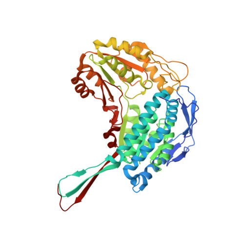
    Analogues of the natural product duocarmycin bearing an indole moiety were shown to bind aldehyde dehydrogenase 1A1 (ALDH1A1) in addition to DNA, while derivatives without the indole solely addressed the ALDH1A1 protein. The molecular mechanism of selective ALDH1A1 inhibition by duocarmycin analogues was unraveled through cocrystallization, mutational studies, and molecular dynamics simulations. The structure of the complex shows the compound embedded in a hydrophobic pocket, where it is stabilized by several crucial π-stacking and van der Waals interactions. This binding mode positions the cyclopropyl electrophile for nucleophilic attack by the noncatalytic residue Cys302, thereby resulting in covalent attachment, steric occlusion of the active site, and inhibition of catalysis. The selectivity of duocarmycin analogues for ALDH1A1 is unique, since only minor alterations in the sequence of closely related protein isoforms restrict compound accessibility.


  • Organizational Affiliation

    Center for Integrated Protein Science Munich CIPSMDepartment of Chemistry, Technische Universität München, Lichtenbergstrasse 4, 85747 Garching (Germany).


Macromolecules
Find similar proteins by:  (by identity cutoff)  |  3D Structure
Entity ID: 1
MoleculeChains Sequence LengthOrganismDetailsImage
RETINAL DEHYDROGENASE 1
A, B
501Ovis ariesMutation(s): 0 
EC: 1.2.1.36
UniProt
Find proteins for P51977 (Ovis aries)
Explore P51977 
Go to UniProtKB:  P51977
Entity Groups  
Sequence Clusters30% Identity50% Identity70% Identity90% Identity95% Identity100% Identity
UniProt GroupP51977
Sequence Annotations
Expand
  • Reference Sequence
Experimental Data & Validation

Experimental Data

  • Method: X-RAY DIFFRACTION
  • Resolution: 1.90 Å
  • R-Value Free: 0.197 
  • R-Value Work: 0.153 
  • R-Value Observed: 0.156 
  • Space Group: P 21 21 2
Unit Cell:
Length ( Å )Angle ( ˚ )
a = 93.556α = 90
b = 150.644β = 90
c = 80.677γ = 90
Software Package:
Software NamePurpose
REFMACrefinement
XDSdata reduction
PHASERphasing

Structure Validation

View Full Validation Report


Ligand Structure Quality Assessment 


Entry History 

Deposition Data

Revision History  (Full details and data files)

  • Version 1.0: 2015-08-26
    Type: Initial release
  • Version 1.1: 2015-09-30
    Changes: Database references
  • Version 1.2: 2015-11-18
    Changes: Database references
  • Version 1.3: 2024-01-10
    Changes: Data collection, Database references, Derived calculations, Other, Refinement description