


Structure of Fesi Protein from Azotobacter Vinelandii
Kabasakal, B., Cotton, C.A.R., Murray, J.W.To be published.
Experimental Data Snapshot
wwPDB Validation 3D Report Full Report

Entity ID: 1 | |||||
|---|---|---|---|---|---|
| Molecule | Chains | Sequence Length | Organism | Details | Image |
| FERREDOXIN, 2FE-2S | 126 | Azotobacter vinelandii | Mutation(s): 0 | 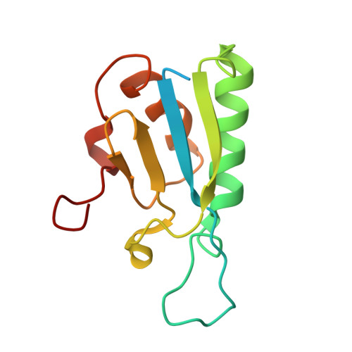 | |
UniProt | |||||
Find proteins for P82802 (Azotobacter vinelandii (strain DJ / ATCC BAA-1303)) Explore P82802 Go to UniProtKB: P82802 | |||||
Entity Groups | |||||
| Sequence Clusters | 30% Identity50% Identity70% Identity90% Identity95% Identity100% Identity | ||||
| UniProt Group | P82802 | ||||
Sequence AnnotationsExpand | |||||
| |||||
| Ligands 1 Unique | |||||
|---|---|---|---|---|---|
| ID | Chains | Name / Formula / InChI Key | 2D Diagram | 3D Interactions | |
| FES Query on FES | C [auth A], D [auth B] | FE2/S2 (INORGANIC) CLUSTER Fe2 S2 NIXDOXVAJZFRNF-UHFFFAOYSA-N |  | ||
| Length ( Å ) | Angle ( ˚ ) |
|---|---|
| a = 39.04 | α = 90 |
| b = 60.1 | β = 109.37 |
| c = 45.51 | γ = 90 |
| Software Name | Purpose |
|---|---|
| REFMAC | refinement |
| XDS | data reduction |
| Aimless | data scaling |
| autoSHARP | phasing |
RCSB PDB (citation) is hosted by
RCSB PDB is a member of the

