5A7F

Comparison of the structure and activity of glycosylated and aglycosylated Human Carboxylesterase 1


Experimental Data Snapshot

  • Method: X-RAY DIFFRACTION
  • Resolution: 1.86 Å
  • R-Value Free: 0.188 
  • R-Value Work: 0.158 
  • R-Value Observed: 0.160 

wwPDB Validation   3D Report Full Report


Ligand Structure Quality Assessment 


This is version 1.3 of the entry. See complete history


Literature

Comparison of the Structure and Activity of Glycosylated and Aglycosylated Human Carboxylesterase 1.

Arena De Souza, V.Scott, D.J.Nettleship, J.E.Rahman, N.Charlton, M.H.Walsh, M.A.Owens, R.J.

(2015) PLoS One 10: 43919

  • DOI: https://doi.org/10.1371/journal.pone.0143919
  • Primary Citation of Related Structures:  
    5A7F, 5A7G, 5A7H

  • PubMed Abstract: 

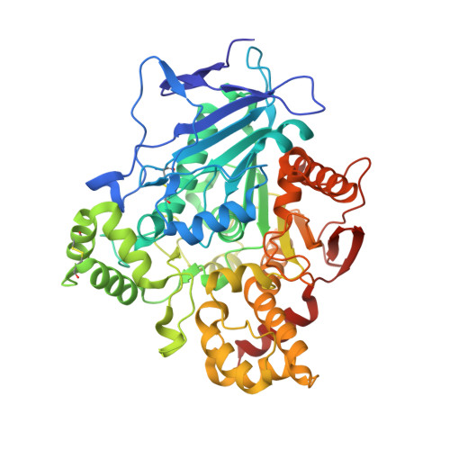
    Human Carboxylesterase 1 (hCES1) is the key liver microsomal enzyme responsible for detoxification and metabolism of a variety of clinical drugs. To analyse the role of the single N-linked glycan on the structure and activity of the enzyme, authentically glycosylated and aglycosylated hCES1, generated by mutating asparagine 79 to glutamine, were produced in human embryonic kidney cells. Purified enzymes were shown to be predominantly trimeric in solution by analytical ultracentrifugation. The purified aglycosylated enzyme was found to be more active than glycosylated hCES1 and analysis of enzyme kinetics revealed that both enzymes exhibit positive cooperativity. Crystal structures of hCES1 a catalytically inactive mutant (S221A) and the aglycosylated enzyme were determined in the absence of any ligand or substrate to high resolutions (1.86 Å, 1.48 Å and 2.01 Å, respectively). Superposition of all three structures showed only minor conformational differences with a root mean square deviations of around 0.5 Å over all Cα positions. Comparison of the active sites of these un-liganded enzymes with the structures of hCES1-ligand complexes showed that side-chains of the catalytic triad were pre-disposed for substrate binding. Overall the results indicate that preventing N-glycosylation of hCES1 does not significantly affect the structure or activity of the enzyme.


  • Organizational Affiliation

    UK OPPF-UK, The Research Complex at Harwell, Rutherford Appleton Laboratory Harwell Oxford, Oxfordshire, United Kingdom.


Macromolecules
Find similar proteins by:  (by identity cutoff)  |  3D Structure
Entity ID: 1
MoleculeChains Sequence LengthOrganismDetailsImage
LIVER CARBOXYLESTERASE 1532Homo sapiensMutation(s): 0 
EC: 3.1.1.1 (PDB Primary Data), 3.1.1.56 (PDB Primary Data)
UniProt & NIH Common Fund Data Resources
Find proteins for P23141 (Homo sapiens)
Explore P23141 
Go to UniProtKB:  P23141
PHAROS:  P23141
GTEx:  ENSG00000198848 
Entity Groups  
Sequence Clusters30% Identity50% Identity70% Identity90% Identity95% Identity100% Identity
UniProt GroupP23141
Sequence Annotations
Expand
  • Reference Sequence
Experimental Data & Validation

Experimental Data

  • Method: X-RAY DIFFRACTION
  • Resolution: 1.86 Å
  • R-Value Free: 0.188 
  • R-Value Work: 0.158 
  • R-Value Observed: 0.160 
  • Space Group: H 3
Unit Cell:
Length ( Å )Angle ( ˚ )
a = 114.715α = 90
b = 114.715β = 90
c = 117.78γ = 120
Software Package:
Software NamePurpose
REFMACrefinement
DENZOdata reduction
SCALEPACKdata scaling
PHASERphasing

Structure Validation

View Full Validation Report


Ligand Structure Quality Assessment 


Entry History 

Deposition Data

Revision History  (Full details and data files)

  • Version 1.0: 2016-01-13
    Type: Initial release
  • Version 1.1: 2019-05-15
    Changes: Data collection, Derived calculations, Experimental preparation, Other
  • Version 1.2: 2020-07-29
    Type: Remediation
    Reason: Carbohydrate remediation
    Changes: Data collection, Derived calculations, Other, Structure summary
  • Version 1.3: 2024-01-10
    Changes: Data collection, Database references, Refinement description, Structure summary