5A49

Crystal structure of the LOTUS domain (aa 139-222) of Drosophila Oskar in C222


Experimental Data Snapshot

  • Method: X-RAY DIFFRACTION
  • Resolution: 2.10 Å
  • R-Value Free: 0.215 
  • R-Value Work: 0.194 
  • R-Value Observed: 0.195 

wwPDB Validation   3D Report Full Report


This is version 1.2 of the entry. See complete history


Literature

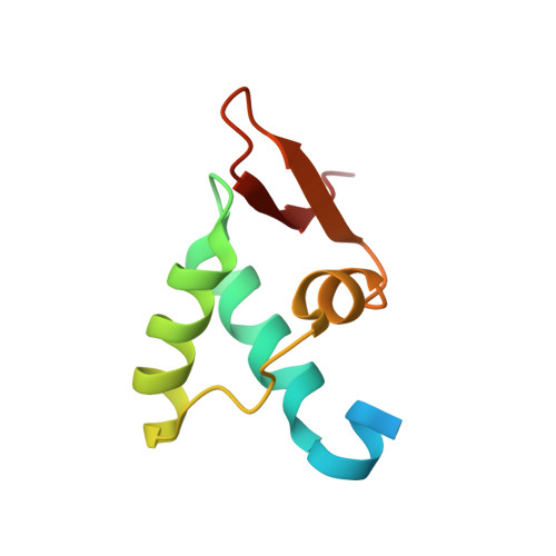
The Crystal Structure of the Drosophila Germline Inducer Oskar Identifies Two Domains with Distinct Vasa Helicase-and RNA-Binding Activities.

Jeske, M.Bordi, M.Glatt, S.Muller, S.Rybin, V.Muller, C.W.Ephrussi, A.

(2015) Cell Rep 12: 587

  • DOI: https://doi.org/10.1016/j.celrep.2015.06.055
  • Primary Citation of Related Structures:  
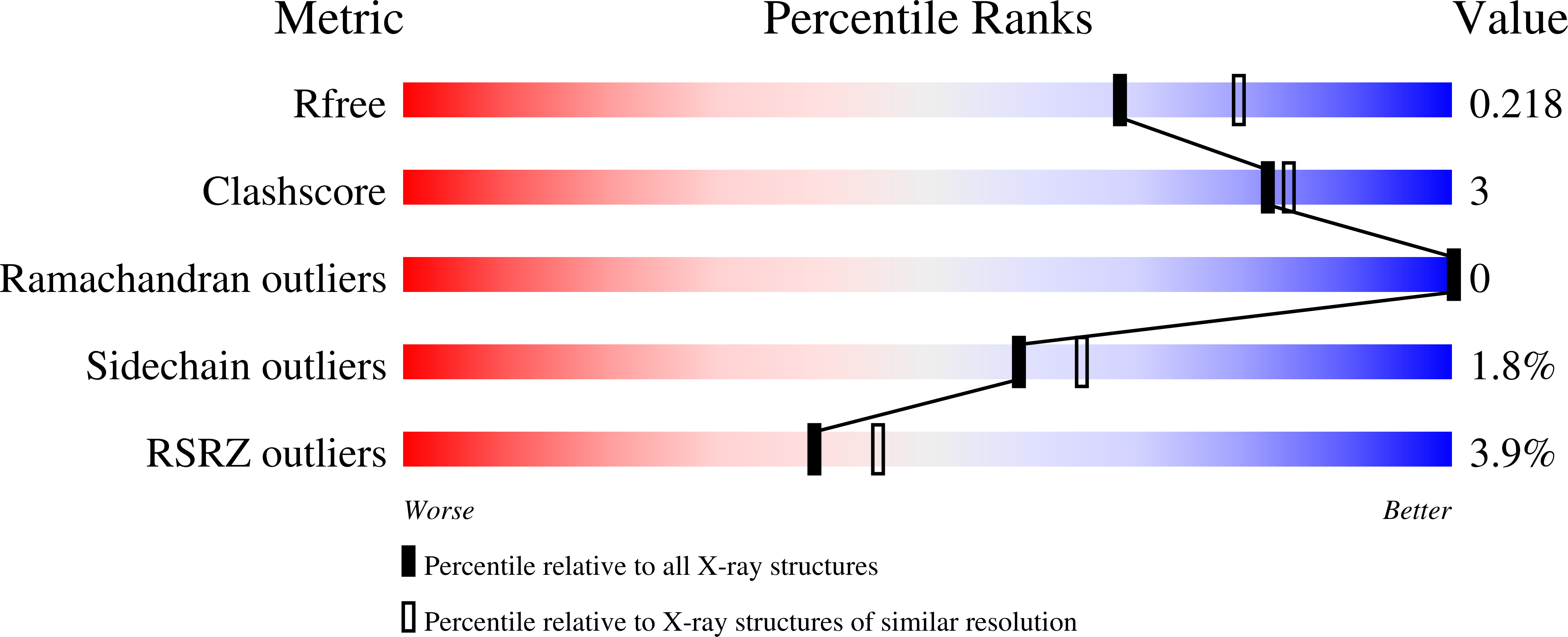
    5A48, 5A49, 5A4A

  • PubMed Abstract: 

    In many animals, the germ plasm segregates germline from soma during early development. Oskar protein is known for its ability to induce germ plasm formation and germ cells in Drosophila. However, the molecular basis of germ plasm formation remains unclear. Here, we show that Oskar is an RNA-binding protein in vivo, crosslinking to nanos, polar granule component, and germ cell-less mRNAs, each of which has a role in germline formation. Furthermore, we present high-resolution crystal structures of the two Oskar domains. RNA-binding maps in vitro to the C-terminal domain, which shows structural similarity to SGNH hydrolases. The highly conserved N-terminal LOTUS domain forms dimers and mediates Oskar interaction with the germline-specific RNA helicase Vasa in vitro. Our findings suggest a dual function of Oskar in RNA and Vasa binding, providing molecular clues to its germ plasm function.


  • Organizational Affiliation

    Developmental Biology Unit, European Molecular Biology Laboratory, 69117 Heidelberg, Germany; Structural and Computational Biology Unit, European Molecular Biology Laboratory, 69117 Heidelberg, Germany.


Macromolecules
Find similar proteins by:  (by identity cutoff)  |  3D Structure
Entity ID: 1
MoleculeChains Sequence LengthOrganismDetailsImage
MATERNAL EFFECT PROTEIN OSKAR
A, B, C, D, E
A, B, C, D, E, F, G, H, I, J
89Drosophila melanogasterMutation(s): 0 
UniProt
Find proteins for P25158 (Drosophila melanogaster)
Explore P25158 
Go to UniProtKB:  P25158
Entity Groups  
Sequence Clusters30% Identity50% Identity70% Identity90% Identity95% Identity100% Identity
UniProt GroupP25158
Sequence Annotations
Expand
  • Reference Sequence
Experimental Data & Validation

Experimental Data

  • Method: X-RAY DIFFRACTION
  • Resolution: 2.10 Å
  • R-Value Free: 0.215 
  • R-Value Work: 0.194 
  • R-Value Observed: 0.195 
  • Space Group: C 2 2 2
Unit Cell:
Length ( Å )Angle ( ˚ )
a = 134.06α = 90
b = 198.63β = 90
c = 89.12γ = 90
Software Package:
Software NamePurpose
PHENIXrefinement

Structure Validation

View Full Validation Report


Entry History 

Deposition Data

Revision History  (Full details and data files)

  • Version 1.0: 2015-07-22
    Type: Initial release
  • Version 1.1: 2015-07-29
    Changes: Database references
  • Version 1.2: 2015-08-12
    Changes: Database references